Webster masterfully translates the abstract complexity of neural circuitry into a tangible spatial reality using physical models. It is a rare example of pedagogical elegance that prioritizes structural clarity over digital gimmickry.
Instala nuestra extensión para buscar dentro de cualquier video al instante
Basal ganglia neuroanatomyAñadido:
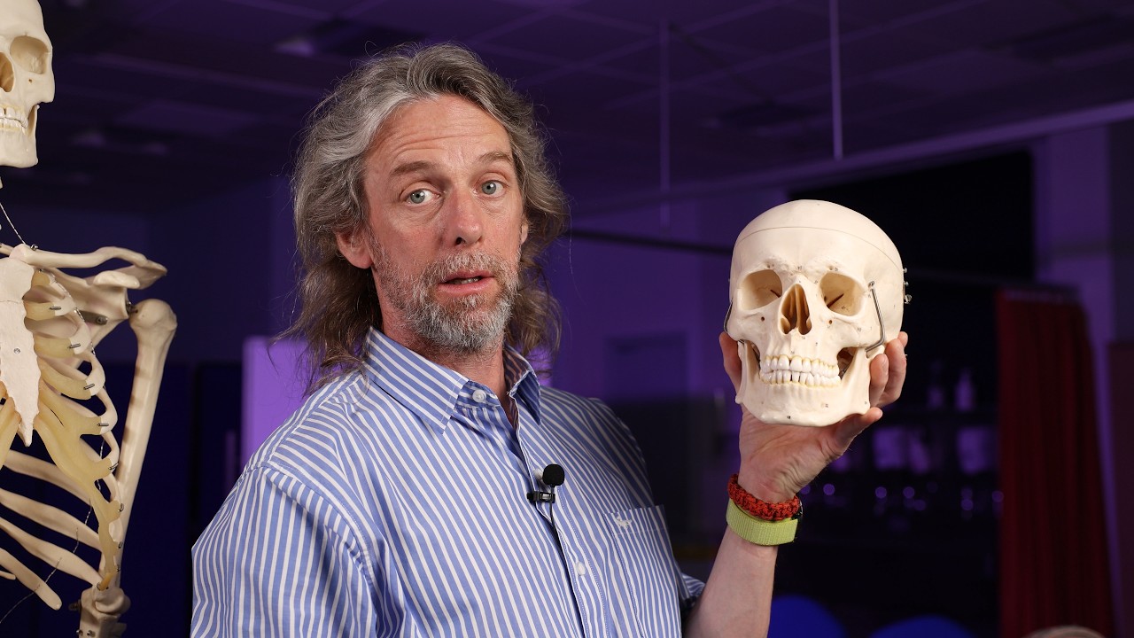
So you want to know about the basil ganglia a or the basil nuclei.
Okay. Um what does an anatomist or maybe a health care professional need to know about the basil ganglia? What we should do is I've got a I've got a bunch of models here is we should see we should find them. We should see where they are and understand them physically as much as we can as well as uh what they're made of. Um and then we'll talk about um how they function, what they do, and by the end of that you'll understand what goes wrong with them when they get injured. Um I I I did a video about the basil gangly back during the COVID times. I was still making videos during during lockdown. I made it in my in my back garden, but my knowledge continues to improve hopefully in my understanding and now we can actually do it with all the models at my disposal. So basil ganglia Basil ganglia. Okay. So, basil here's the here's the brain. So, the cerebral hemispheres down there is the brain stem. There's the cerebellum. So, the base of the brain, the base of the cerebrum is in there somewhere, right?
That's what the basil bit refers to.
It's deep. So we've got to go hunting for the parts of the basil ganglia.
Ganglen. A ganglen is a collection of neuron cell bodies in the peripheral nervous system. That is you've got your you got your neuron. It's got this nice big cell body and then it sends its axons out off at a distance somewhere.
So where you have those neuron cell bodies collected together makes a little bit of a lump. That's a ganglion. That means it's gray matter because white matter would be mileelinated axons.
Right? This is gray matters. This is where you have connections. This is where where you have circuits where you have complexity. Now a collection of neuron cell bodies in the central nervous system should be called a nucleus. So the contemporary name for these structures really is the are the basil nuclei rather than the basil ganglia. But we're kind of stuck with basil ganglia because that's what everybody calls them. Anyway, you'll hear both. The basil ganglia are not one structure. There are a number of structures deep in the base of the brain. So they've got separate structures. They got separate names.
They all get grouped together as the basil ganglia. And they all get grouped together in other ways as well. So the terminology can be a bit complicated.
Let's first of all uh figure out where we are. Okay. So you see you see where we are. We're in the head. Um, and if we slice our way down and slice again and slice again, now we can see the the lateral ventricles. Need to go a little bit deeper.
Okay. Now, what level are we at? Huh?
There's the eyes. So, there's the eyebrows.
So, now we can see some gray matter in the base of the brain. So again these shapes here are the lateral ventricles but these these are bit of gray matter back here we've got the phalamus the phalamus on either side all the structures I'm going to talk about we have one on either side they are symmetrical as it were so we have the phalamus and then these patches here and here and here these are some of the basil ganglia this looks like a lens this is the lentapform nucleus and actually within the lentapform nucleus the outer part is called the putwamin And the inner part is called the globus paladus. And then actually the globus paladus is broken up into an internal part and an external part.
And then here we've got the head of the cordate nucleus. Cordate. Well, that means corder is Latin. It means tail. So the cordate nucleus, this is the head of the cordate nucleus. It actually loops over and around. So it has this long tail type structure. So, lentifiform nucleus, cordate nucleus, phalamus. So, the phalamus isn't part of the uh basil ganglia, but it's a really good landmark. The phalamus um carries sensory information back up to the cortex. Well, that's part of the story anyway. We'll see there's more to that.
Okay, go deep a deeper layer. Now, we can see parts of the lateral ventricles back here. And again, we can still see that lentifiform nucleus putin globus paladus. We can actually see some indications here of the separate parts cordate nucleus and the phalamus.
Now um the subthalamic nuclei are subthalamus. They are deep. They are inferior to the phalamus and they're between the phalamus and the brain stem.
But I'm not going to see them in this plane of section. I don't think I'm going to be able to show them in any way. But inferior to the phalamus are the subthalamic nuclei. So if we go down to the next layer, we've actually gone down to the midbrain. So if the brain stem is made up of the medulla longata, then the pawns, then the midbrain, we're looking at the midbrain. Here we can see these two stalks. These are the cerebral podunks which are going to carry tracts um to and from the cerebral hemispheres.
Um, and then we've got various midbrain stuff in there. The level we're at now, there's the optic kayazm. So, there are the eyes.
We can see we're within the top of the orbits there. Uh, and we've got the um internal uh corroted arteries on either side. So, that's the level that we're at. If we go any deeper, now we're getting further down the the brain stem and further away from what we're interested in. Now in the brain stem if we're talking about the basil ganglia another component of the basil ganglia is here in the midbrain and it's this dark patch here. So if these are the cerebral podunkils, these dark patches on the cerebral podunk side, the anterior side, these are the substantia The substantia some of the cells in here are producing a neuromelanin, a form of melanin, the thing that gives you the pigment in your skin. Um, so that that's why we see the pigment. This is literally a dark patch, substantia the dark substance.
And these cells here, there are two parts to the substantionigra. There's the par reticularis and there's the par compactor.
Um, and these are projecting up to those other parts of the basil gangula that we looked at. So the substantia is in the midbrain.
So that's one way of looking at the basil ganglia. And if you're looking at radiological images, uh, MRI transverse sections for example, you can see those same structures in that same plane of section there. And again, you can see the putwamin, the globus paladus, the cord nucleus, and then the phalamus.
Again, the phalamus is not part of the basil ganglia, but it is intimately related.
That is a heavy model. Let's look at this again from another perspective before we go and take the brain apart which is going to be the trickiest bit.
So here we go half ahead. So midsagittal section and um here uh this little depression here is the third ventricle and the wall of the third ventricle that kind of oval shape that we can see there that's the phalamus. So the medial edge of the phalamus there the hypothalamus is down here.
Um, and if we look at the brain stem, there's the midbrain. So, if we have the pawns is the obvious nice bulgy curved bit. The midbrain is the bit of the brain stem superior to the pawns or between the pawns and where we see the phalamus up here. So, the midbrain is kind of it's kind of going up like like that. You know, a lot of it's been cut away. Anyway, stop talking, Sam. Keep it straight forward. Um, there is the midbrain. And we can see the cerebral aqueduct passing through it caliculi on the posterior surface um and what have you.
If this is the midbrain and this is the phalamus then the subthalamic nucleus is going to be in there somewhere right? It's going to be inferior to the phalamus and superior to the brain stem. It's going to be in there subthalamic nucleus if that helps.
So it's going to be superior to where we find the substantia.
Now this here that is the septum palucidum. Septum being a divider.
Pucidum is kind of translucent. Um and you've got those lateral ventricles we were looking at are kind of on the other side of that. Now there's another nucleus called the nucleus acumb. And the nucleus encumbent sits inferior to the head of the cordate nucleus. and kind of kind of leans up against the septum palucidum. So that's kind of just on the other side of that. Not sure if that's going to help, but you can see our layout from another perspective.
Midbrain, phalamus, and then the basil ganglia are they're around here, but in there. Okay, time to take the brain apart.
So, you sure you want to take this apart? Because it doesn't go back together very easily.
All right, let's take the cerebellum off so we can see the brain stem. To be honest, this is likely to fall apart.
Um, let's take let's take this off. So, a big part of the cerebrum.
And now we can see the insula cortex hidden away in there. This clear structure here, that's that lateral ventricle again.
Uh, okay. Let's take off the other side.
Um, let's take off the frontal lobe. So, you see how we're getting we're dissecting away. We're getting deeper and deeper, right? Um, let me Okay, let me take that off. Now, if I spin this around, so we're looking at the left side of the brain. If I peel off the insular cortex, we can see some red.
Um so the model here is painted red to indicate that is the putwamin. So the outer part of that lentififor nucleus the putwamin.
Um I need to take off the um lateral ventricle. So I think I need to take off the rest of the brain. Okay.
So now we've got we've just got brain stem basil ganglia and the phalamus is in there as well. Let me take off the um the lateral ventricles. Oh, so of course they're in the middle as well, aren't they? It's the whole ventricular system as a cast. So that means that we know where the phalamus is because we said that the phalamus is um up against that that third ventricle there.
So now I've taken this apart. Do you see what I mean? Now we've dissected it. It kind of gets a bit more confusing. But if I dissect that and open it up, that's the phalamus there because we said there's the pawns, there's the midbrain, there's the third ventricle.
So the rugby ball shape there up against the third ventricle is the phalamus and there's the hypothalamus. Right? Let me put that back together just so we've got a bit more of a structure. Do you see where we are? So medulla pawns midbrain is is up in there. So, as I was saying, there's the putwamin. So, if you remember our cross-section through here, uh, we saw the head of the cordate nucleus up there as well. So, that there, that's the cordate nucleus.
There's its tail looping around like that. So, that's the cordate nucleus and that's the putwamin.
Okay? So that nuclear circumbent is up in there inferior to the head of the cordate nucleus. And if that's the putwamin that means I would need to take this off to see the uh globus paladus. So if I take this off um it's in there.
So that bulge there that's the globus paladus with its internal part and its external part.
Uh these here are white matter tracks running to and from various structures.
Right? So the basil ganglia are the putwamin, the cordate nucleus, the globus paladus external segment and the globus paladus internal segment, the nucleus acumbent, the subthalamic nucleus and the substantia Right, there's more.
If you cut sections through the brain, when you cut sections through the basil ganglia, it can look a bit stripey. Um, the word we use for stripey things in anatomy is uh striated.
Um, so we have something called the striatum. Now the word striatum is actually linking together three parts of the basil ganglia the parts that we've talked about. So the striatum if you use the term striatum it refers to the nucleus circumbent the cordate nucleus and the putwamin and you may also hear about the dorsal striatum and the vententral striatum.
Well, more dorsally we have the cord a nucleus and the putwamin and more ventrally we have the nucleus circumbent and something called the alfactory tubicle that I am not going to point at.
So that's if you hear about dorsal stryatum and vententral stratum those are terms that are linking together these structures that we've talked about and when we're going to talk about the functions of the basil ganglia we are going to talk about the stryatum. So when I say strriatum I am lump I am lumping together cordate nucleus the putwutamin and the nucleus acumbance.
Okay H so what are the functions of the basil ganglia?
Most importantly movement in a word but there's more. Um the basal ganglia is is is it's quite comp it's very complex. it's not entirely understood. Um, so however, so whenever it's simplified and distilled, you lose some of the finesse. But I mean, I'm over really simplifying if I say movement, but movement. Um, there are inhibitory circuits within the basil ganglia. So it can be helpful to think of the basil ganglia circuits as um inhibiting unwanted movement. And for movement to occur, you have to inhibit that inhibition.
That can be a helpful way of thinking about this. Um, now the striatum has inputs, well, lots of inputs from many regions of the brain, but it has inputs from the cerebral cortex. And the subthalamic nucleus has inputs from the cerebral cortex.
And then the basil ganglia there's a whole bunch of there are a whole bunch of circuits within the basil ganglia but then it has outputs from the um substantia par reticularis and the globus paladus internal segments and those outputs go to the phalamus and affect phalamus function. So there are some nuclei in the phalamus that are involved in movement involved in motor outputs that go back to the cerebral cortex. So we have those particular inputs and those particular outputs and then circuits within the basil ganglia.
There are three pathways that we can talk about that have been elucidated that have been figured out that describe how these parts of the basil ganglia uh interact with movement.
um we have the direct pathway, the indirect pathway and the hyperdirect pathway.
And let me just review what I just said.
So um the stryatum has a whole has lots of inputs from the cerebral cortex and those inputs are uh glutamate mediated.
Glutamate is the neurotransmitter.
uh and then the outputs from the basil ganglia to the phalamus those are from the par reticularis of the substantia and from the internal segment of the globus paladus those output to the phalamus to the ventro anterior and vententralateral nuclei in the phalamus that are responsible for motor outputs and movement bits of the phalamus the pars compactor part of the substantia projects up to parts of the basil ganglia and modulates the activity of the circuits between those inputs and the outputs. The circuits between the intrinsic nuclei largely within the within the basil ganglia and those outputs from the past compact part of the substantia are dopamine mediated. So we've got glutamate, dopamine and GABA getting involved here.
Now with the direct pathway you want to initiate a voluntary movement and the cortex sends excitatory inputs into the striatum.
And the striatum then sends inhibitory inputs and inhibits the um the neurons in the internal segment of the globos paladus and the uh the pars reticularis of the substantia So this the the striatum is inhibiting the inhibition of those outputs onto the phalamus.
The nuclei in the phalamus can now activate. They can now send excitatory signals back up to the cortex and initiate a movement. The direct pathway is the way in which you initiate a voluntary movement.
Right?
the indirect pathway.
Again, excitatory signals are sent into the stryatum.
Um, but the stryatum sends inhibitory sing signals to the globus paladus externus.
Now the globus paladus external segment would normally be sending inhibitory signals to globus paladus internal segment the pareticularis of the substantionigra and to the subthalamic nucleus.
So these new excitatory inputs are inhibiting the globus paladus external segment. So it is now no longer inhibiting the subthalamic nucleus and now the subthalamic nucleus can send excitatory outputs to the globus paladus internal segment and the pareticularis of the substantia and now those can inhibit the motor nuclei of the phalamus and prevent unwanted movement. So you can think of the direct pathway as initiating movement and the indirect pathway of stopping movement. But it is now understood that both of those pathways are working together at the same time such that you are making the movements that you want to make but you are in you are inhibiting the movements that you do not want to make. And this is crucial to precise movements, right?
So the direct pathway and the indirect pathways uh inhibiting parts of the phalamus and inhibiting the inhibition of parts of the phalamus. These circuits are giving control over movement that control that we take for granted. What about the hyperdirect pathway then?
Well, the hyperdirect pathway is so named because those excitatory outputs from the cortex completely bypass the stryatum and go straight to the subthalamic nucleus.
So those neurons in the subthalamic nucleus then send excitatory outputs to the pareticularis of the pareticularis of the substantia and the internal segments of the globus paladus telling the neurons there to inhibit those motor nuclei in the phalamus stopping unwanted movement. So the hyperdirect pathway supports the indirect pathway in preventing unwanted movement.
How are you doing?
Um we're learning that the basil ganglia are involved in more than just movement.
There are links to the prefrontal cortex. It's involved in decision making, planning, reward, motivation, possibly uh aspects of memory and learning, um behavior, um and a whole bunch of things nobody knows about yet because we haven't learned it. We haven't understood it. We haven't found it out yet. But for most of us, the basil ganglia are involved in movement and other functions. So from what we've discussed hopefully it's pretty clear why damage to structures of the basil ganglia leads to movement disorders such as tremor. uh Parkinson's disease um is a disease in which the cells of the par compactor of the substantia die slowly over time the par compactor those neurons uh are sending axons up into the striatum and they release dopamine and the dopamine there um it it aids in activation of the direct pathway and inactivation of the indirect pathway. So those neurons are important in in initiating a voluntary movement and that is a a key sign in Parkinson's is the difficulty in in initiating a new movement. But that said, Parkinson there's more to Parkinson's than just that and not all Parkinson's um exhibit the same signs and symptoms. But the circuits, the structures that we've described in the basil ganglia hopefully help you understand what we see in those movement disorders um where parts of the basil ganglia have been injured. And there is much more to learn and there's much more to read. And if you want to find out more about the neuroscience, go out there and read. I'll maybe put a couple of links in the description to this video. But there's there's, you know, I'm focused on the neuro anatomy, the names of the lumpy bumpy bits. But there's a lot more detail to this if you want to go and discover it. All right, I hope that was useful. I hope that made some sort of sense.
Um, but the basil ganglia, would you have guessed it? Me talking about the basil ganglia. Speak to you uh next time.
Videos Relacionados

Recovery pronouns. Neuroplasticity & practical neuroscience tips to help recover from pain & fatigue
Fantasticneuroplastic
907 views•2026-05-31

No Eyes, No Darkness? 👀😱
Huwatif
630 views•2026-06-02

I Saw the Thing Crash. Then I Lost Hours | Beyond Black Budget
BeyondBlackBudget
148 views•2026-05-30

Your Brain Is Actively Deleting Your Childhood Memories! 🧠🗑️ #Shorts #Anatomy #DidYouKnow
voiceless2345
225 views•2026-06-01
Neuroanatomy of smell (olfaction)
SamWebster
644 views•2026-05-28

What are you looking at
SuperStaticPro
1K views•2026-05-31

Size Illusion
WTFactt_t
1K views•2026-06-03
Why Trauma Doesn’t Just 'Go Away'
historyofsimplethings
1K views•2026-05-28


