This analysis offers a rigorous neurobiological validation for insomnia, moving far beyond the usual superficial sleep advice. However, feeding a hundred facts to an exhausted mind is a brilliant irony that risks fueling the very hyperarousal it seeks to explain.
Install our extension to search inside any video instantly.
Why Your Brain Can't Stop: 100 Science Facts for an Exhausted Mind - No AdsAdded:
I'm grateful you found your way here tonight. You are exhausted.
And yet here you are, still thinking, still processing, still unable to fully stop. That gap between what your body needs and what your mind allows is exactly what tonight is about. Welcome to sleepy mind science.
Tonight we are going to move through 100 science facts about brain hyperarousal.
The state where your mind refuses to quiet down no matter how tired you are will travel from neuroscience to evolutionary biology, from stress hormones to sleep chemistry.
And somewhere in that journey, your mind will let go. Before we begin, if this channel has ever helped you find sleep on a night when nothing else worked, consider subscribing.
Every person who finds this channel through your support is another exhausted mind that gets a chance to finally rest. We grow one sleepy soul at a time. You do not need to solve anything tonight. Your thoughts are allowed to exist without being finished.
Let your body feel the weight of the bed beneath you. Like a dark room after the last light goes out, still unheld.
And finally, only yours. You can rest here. Close your eyes and breathe deeply. You're resting where a storm of thoughts finally meets silence. Let your mind soften like a signal fading. And now we begin.
Brain hyperarousal is a state of excessive mental and physiological activation that persists even when the body is physically exhausted. During hyperarousal, the brain's threat detection circuits remain switched on, firing continuously, even in the absence of any real danger.
Researchers at the National Institute of Mental Health have documented elevated metabolic activity in the brains of people with chronic insomnia compared to those who sleep normally even during the night when sleep should be occurring. The brain is in effect running a full alarm cycle through hours that were biologically designed for repair and restoration.
What makes this state so confounding is that the very exhaustion it produces does not switch it off. It can intensify it. The amygdala, an armond-shaped region deep in the brain's temporal lobe, plays a central role in hyperarousal.
This structure is the brain's primary threat evaluation center, scanning incoming sensory information and emotional memory for anything that requires a defensive response in people who experience chronic hyperarousal.
The amygdala shows heightened baseline activity even during rest. According to neuroiming research published in the journal Sleep, this means the brain is not simply responding to stress when it appears. It has recalibrated its resting state to assume that stress is always present. The amygdala does not distinguish between a physical predator and an unresolved email. Both receive the same urgency signal. Cortisol, the body's primary stress hormone, follows a predictable daily rhythm in healthy sleep patterns. It peaks sharply in the early morning to support waking alertness, then declines steadily through the day, reaching its lowest point in the late evening, a biological signal that prepares the brain and body for sleep.
In people experiencing chronic hyperarousal, this rhythm is flattened or disrupted.
Evening cortisol levels remain elevated rather than declining, keeping the nervous system in a state of readiness precisely when it should be entering the quiet of pre-leep deactivation.
This hormonal pattern is one of the most consistent biological markers researchers have found across people who report lying awake despite exhaustion.
The locus coilus is a tiny cluster of neurons in the brain stem that produces nearly all of the brain's norepinephrine, a neurotransmitter associated with arousal, alertness, and threat response. When the locus corius is active, the brain is tuned for rapid detection and response. Sleep requires this system to quiet down substantially.
In animal studies, locus corulius neurons show near complete silence during nonm sleep. In people with hyperarousal, this silencing appears to be incomplete or delayed. The neurons continue firing at low but sleepd disrupting rates well into the night. The system is in an evolutionary sense doing exactly what it was built to do, keeping the organism alert in an environment it perceives as unsafe.
The preffrontal cortex, the brain region most associated with rational thought, planning, and the regulation of emotion, serves as a break on the amygdala's alarm system. Under normal conditions, the preffrontal cortex modulates amygdala activity, dampening fear responses once they have served their purpose. Sleep deprivation significantly impairs this regulatory connection. Research from the University of California, Berkeley found that after just one night of sleep loss, amygdala reactivity to negative stimuli increased by 60%.
While functional connectivity between the prefrontal cortex and amydala weakened. This means that the more sleepd deprived you become, the harder it is for the rational brain to calm the alarmed brain, creating a cycle that worsens with each restless night.
Adenosine is a chemical byproduct of neural activity that accumulates in the brain throughout the day. As adenosine builds up, it binds to receptors in the brain that progressively inhibit wakefulness and create the sensation of sleepiness, a process researchers call sleep pressure. After approximately 16 hours of wakefulness, adenosine levels are high enough to drive strong sleep pressure in most people. However, in hyperarousal states, the activating systems of the brain driven by cortisol, norepinephrine, and other arousalpromoting neurochemicals can override this pressure. The body is sending a clear biological signal to sleep. The threat detection system is overriding it. Adenosine pressure accumulates regardless.
which is why people in hyperarousal states feel profoundly exhausted but cannot stop. The hypothalamus contains a region called the vententralateral preoptic nucleus often abbreviated VLPO which functions as the brain's primary sleep initiation center. When the VLPO becomes active, it sends inhibitory signals to the arousalpromoting systems of the brain stem and hypothalamus, effectively switching off wakefulness.
The relationship between sleep and arousal systems in the brain has been described as a flip-flop switch. One side must be clearly dominant for a stable state to be maintained. In hyperarousal, the arousal side of this switch remains dominant even when adenosine pressure and circadian cues are pushing for sleep. The V LPO cannot gain enough activation to flip the system into stable sleep. The brain oscillates at the edge without fully crossing over.
Hypervigilance, the state of sustained high alertness to potential threats, is behaviorally distinct from ordinary wakefulness, but produces many of the same neurological signatures as hyperarousal.
People in hypervigilant states show elevated startle responses, heightened sensitivity to sound and light, difficulty filtering irrelevant stimuli, and a persistent sense that something requires their attention.
Research using polymnography has shown that hypervigilant individuals have measurably increased highfrequency brain activity during sleep itself particularly in the beta frequency range. This suggests the brain is not fully disengaging from monitoring activity even during sleep stages that should be characterized by slow low frequency waves.
The body's autonomic nervous system is divided into two primary branches. The sympathetic system which drives the fightor-flight response and the parasympathetic system which supports rest, digestion and recovery. Healthy sleep requires a shift toward parasympathetic dominance as evening progresses.
Heart rate variability, a measure of the variation in time between heartbeats, serves as an accessible proxy for this balance.
Higher heart rate variability is associated with greater parasympathetic activity and is considered a marker of physiological readiness for sleep and recovery. In people experiencing chronic hyperarousal, evening heart rate variability is often reduced, suggesting that the sympathetic system remains activated through the hours that should be transitioning toward rest.
Rumination, the repetitive passive focus on distressing thoughts and feelings, is one of the most consistent cognitive features of hyperarousal related sleep difficulty.
Unlike productive problem solving, rumination does not move toward resolution.
It cycles through the same material repeatedly, drawing from the same pool of worry or regret without generating new information or decision outcomes.
Neuroiming research has identified heightened activity in the default mode network.
The brain's self-referential processing circuit during periods of rumination.
This network, which is normally supposed to quiet down during sleep preparation, remains highly active in people who report racing thoughts at bedtime. The brain is in effect reviewing its own concerns as if reviewing them more thoroughly would lead to resolution. The default mode network or D MN is a set of interconnected brain regions including the medial preffrontal cortex, posterior singulate cortex and hippocampus that becomes most active during internally directed thought self-reflection imagining future scenarios and reviewing past events. It is sometimes called the brain's resting state network, but this name is misleading. It is not a resting state so much as a self-focused processing state.
During healthy pre-sleep transitions, DMN activity gradually declines as the phalamus begins to gate sensory input and the cortex shifts towards slower oscillations.
In chronic hyperarousal, DMN deactivation is incomplete or delayed, keeping the brain focused inward on unresolved concerns rather than releasing into the more diffuse neural activity that characterizes approaching sleep. Interception is the brain's capacity to sense the internal state of the body, heart rate, breathing rhythm, temperature, muscular tension, and dozens of other physiological signals. The insular cortex, a region tucked within the lateral sulcus of the brain, is the primary processing hub for introsceptive information. in hyperarousal states.
Research has found that insular cortex activity is elevated and that people show heightened sensitivity to internal body signals, particularly those associated with threat, such as increased heart rate or shallow breathing. This creates a feedback loop. The brain's arousal state produces physical tension. The heightened interception detects that tension as a signal of threat or danger and the amygdala responds by increasing arousal. Further, thermal regulation plays a less discussed but scientifically significant role in sleep initiation.
Core body temperature must fall by approximately 1 to 1/2° C for sleep to begin efficiently. This temperature drop is actively orchestrated by the brain through vasoddilation.
The widening of blood vessels in the hands and feet which releases heat and allows core temperature to decline in people with hyperarousal.
This thermore regulatory process is disrupted.
Research published in the journal Psychossematic Medicine found that people with chronic insomnia showed impaired pre-sleep core body temperature decline compared to good sleepers, suggesting that the arousal state interferes with one of the most fundamental physical preconditions for sleep onset. Gamma aminobuteric acid known as GABA is the brain's primary inhibitory neurotransmitter.
It functions as a neurological break, reducing neural excitability across broad circuits of the brain and facilitating the transition from high activity arousal states to the quieter states required for sleep. Many widely used sleep medications work by enhancing GABA activity at specific receptor sites.
Research using magnetic resonance spectroscopy has found that people with primary insomnia show reduced GABA concentrations in parts of the brain associated with arousal regulation, suggesting that the chemical system most responsible for quieting neural activity may be less robustly active in people who experience chronic difficulty falling asleep.
Sleep is not a single uniform state. It is a sequence of distinct stages each with characteristic patterns of brain activity, eye movement, muscle tone, and hormonal activity. The transition from wakefulness into sleep begins with stage one nonrem sleep, a light state in which alpha brain waves give way to slower theta waves and the muscles begin to relax. In normal sleep transitions, this stage lasts only a few minutes. In hyperarousal states, stage one can be prolonged significantly with the brain repeatedly generating activity that pulls it back toward wakefulness before fully entering the deeper, more restorative stages. The sleep architecture of people with chronic hyperarousal often shows fragmented stage 1 entries, fewer slowwave sleep periods, and more nighttime awakenings.
The hippocampus, the brain's primary memory consolidation center, is acutely sensitive to stress and arousal related neurochemicals.
Elevated cortisol in particular has well doumented effects on hippocample function and structure.
Short-term cortisol elevation enhances certain types of memory formation particularly for emotionally significant events.
An evolutionary feature designed to help organisms remember dangerous situations.
But chronic cortisol elevation suppresses hippocample neurogenesis and impairs the hippocampus's ability to process and contextually situate memories. In the context of hyperarousal related sleep difficulty, this creates a particular problem. The memories and worries the brain is cycling through at night may be processed less effectively, reducing the brain's capacity to emotionally regulate and contextualize them. Emotional memory reconsolidation, the process by which stored emotional memories are updated and reprocessed when recalled, is one of the functions that sleep appears to support. Research by neuroscientist Matthew Walker and colleagues at the University of California, Berkeley suggests that sleep in particular serves as a form of overnight emotional processing, allowing the brain to replay emotionally charged memories in a neurochemical environment low in stress related norepinephrine.
This reduced norepinephrine replay may allow the emotional intensity of memories to be gradually diminished over time. In hyperarousal states, the reduction of norepinephrine during is incomplete, potentially limiting the extent to which sleep can perform this emotional regulation function. Chronic hyperarousal is associated with a persistent low-grade activation of the HPA axis. the hypothalamic pituitary adrenal axis which coordinates the body's long-term stress response. The HPA axis operates through a feedback loop. The hypothalamus signals the pituitary gland which signals the adrenal glands to release cortisol. Rising cortisol then signals back to the hypothalamus and pituitary to reduce further production.
In chronic stress, this negative feedback loop can become disregulated with the HPA axis failing to fully suppress itself even after the stressor has passed. The result is a body that continues producing stress hormones in the absence of acute stress, maintaining the physiological conditions of threat readiness around the clock. The concept of alostostatic load describes the cumulative biological cost of chronic stress exposure on the body and brain.
Developed by neuroscientist Bruce Mchuan, the concept refers to the wear and tear that results from repeatedly activating and failing to fully deactivate stress response systems. High alosatic load is associated with elevated inflammation.
disrupted hormonal rhythms, impaired immune function, and reduced cognitive flexibility.
All of which have been independently linked to sleep disruption. In this framework, chronic hyperarousal is not simply a sleep problem. It is one expression of a broader physiological state in which the body's adaptive systems have been chronically overloaded and have begun to create costs across multiple biological systems simultaneously.
Inflammatory markers particularly interlucan 6 and tumor necrosis factor alpha are elevated in people who report chronically poor sleep quality. Inflammation and the immune response system have complex birectional relationships with sleep. The brain actively uses sleep time to regulate immune function and immune activation can in turn disrupt sleep architecture.
Research has found that even modest sleep restriction increases circulating inflammatory markers within days. In the context of hyperarousal, this creates another reinforcing cycle.
The arousal state disrupts sleep.
Inadequate sleep elevates inflammation.
Elevated inflammation further sensitizes the nervous system to stress signals.
And a more sensitized nervous system generates more hyperarousal.
Sensory gating is the brain's ability to filter and suppress the processing of repetitive or irrelevant sensory stimuli, allowing a person to habituate to background noise, ambient light, or minor physical discomfort and shift attention away from them. Healthy sleep onset depends substantially on sensory gating. The brain must progressively reduce its engagement with environmental stimuli as sleep approaches.
Research using auditory evoked potentials has found that people with insomnia and hyperarousal show reduced sensory gating compared to normal sleepers. Their brains continue to fully process sensory input that should be filtered out, keeping the brain in an active monitoring posture rather than a passive one. The parasympathetic nervous system's primary communication pathway to the visceral organs runs through the vagus nerve. The longest cranial nerve in the body extending from the brain stem through the chest and abdomen.
Vagal tone a measure of the activity level of this nerve is closely associated with parasympathetic activity.
emotional regulation and the capacity for relaxation and social engagement.
Higher vagal tone is consistently associated with better sleep quality, greater emotional resilience and lower inflammatory profiles.
In people with chronic hyperarousal, veagal tone is often reduced, meaning the nervous system's capacity to actively shift into rest and recovery mode is compromised by the same chronic activation that produces the hyperarousal in the first place.
Sleep spindles are brief bursts of synchronized neural activity appearing as distinctive spindle-shaped patterns on EEG recordings that occur primarily during stage 2 nonrem sleep. They are generated through oscillating activity between the phalamus and the cortex and are thought to serve several protective functions including suppressing the processing of external stimuli during sleep and supporting memory consolidation.
Research has found that people with insomnia and hyperarousal tend to generate fewer sleep spindles per hour of stage 2 sleep than people without sleep difficulties.
This reduced spindle activity may be one mechanism through which the hyperaroused brain remains more reactive to environmental stimuli and wakes more easily during the night. The phalamus acts as the brain's central relay station, routing sensory information from the body and external environment to appropriate cortical processing areas. During sleep, the phalamus performs an additional function. It enters a mode of phalamocortical gating in which it actively reduces the flow of sensory input to the cortex creating a kind of neurological wall between the sleeping brain and the environment.
This gating is essential for maintaining sleep in the presence of normal background noise and movement. In hyperarousal states, theamic gating appears to be less complete with sensory signals reaching the cortex more readily during sleep.
One possible explanation for why people with chronic hyperarousal report being more easily awakened by sounds or movement during the night. The phenomenon of paradoxical insomnia in which individuals perceive themselves as having slept very little or not at all while polyomnographic recordings show substantial sleep offers a window into the subjective experience of hyperarousal during sleep. Research suggests that people who experience this mismatch between perceived and objective sleep show elevated highfrequency brain activity during sleep stages that should be dominated by slow wave oscillations.
The brain is technically asleep by most standard measures but a part of it remains active in the monitoring self-aware mode associated with wakefulness.
The result is sleep that does not feel like sleep. Rest that is not experienced as rest because a portion of the brain continues to watch even while the rest of it has gone quiet. Notice the weight of your body where it meets the surface beneath you. Feel how much is already being held without effort, without your help. Your lungs are breathing. Your heart is beating. Your cells are doing their work in the dark. You've been carrying a mind that wouldn't stop tonight. That is not a flaw. That is a system designed over millions of years to keep you safe. It has been doing its job with extraordinary dedication.
And now for a little while, you can tell it I am safe. There is nothing here that needs your protection tonight. Let the storm of neurons quiet. One frequency at a time. You are still here and you are allowed to rest. Hyper arousal associated with sleep difficulty is not identical to generalized anxiety.
Although the two conditions frequently co-occur and share neurobiological features, researchers distinguish between state arousal, a temporary activation triggered by specific stressors, and trait arousal, a stable baseline disposition toward heightened nervous system activity that persists across contexts and over time.
People with chronic insomnia, characterized by hyperarousal, often show trait level elevations in physiological activation that are present even in the middle of the day, not only at bedtime. This trait level finding suggests that for many people, the difficulty falling asleep is a symptom of a broader nervous system orientation rather than a problem specific to the bed or the bedroom.
childhood adversity, including experiences of neglect, abuse, chronic family conflict, or early loss, has well doumented effects on the development of the stress response systems that underly adult hyperarousal.
Research in developmental neuroscience has shown that adverse early experiences can alter the set point of the HPA axis.
increase baseline amydala reactivity and reduce the density of glucocorticoid receptors in the hippocampus.
All of which contribute to a nervous system calibrated for ongoing threat detection. These early calibrations are not defects in the person but adaptations to the environment in which that person developed. The brain learned accurately that vigilance was necessary. Unlearning that lesson is a biological as much as a psychological process.
The concept of alostostasis coined by physiologists Peter Sterling and Joseph Ay extends beyond the older concept of homeostasis by recognizing that the body does not simply maintain fixed biological set points but actively anticipates and adjusts to anticipated demands. In alistasis, the brain uses predictive models of the environment to prepare the body's stress systems in advance.
This anticipatory quality means that hyperarousal can be triggered not only by current threats, but by predicted ones, by learned patterns, by contextual quiz, by the approach of bedtime itself. For many people who experience difficulty sleeping, the bedroom and the act of lying down have become powerful conditioned cues for nervous system activation because the brain has learned to associate that context with the experience of wakefulness and distress.
Stimulus control, the association between environmental cues and behavioral states is one of the most robustly studied mechanisms in sleep science. Classical conditioning research demonstrated that animals and humans readily form associations between neutral environmental stimuli and physiological states when those stimuli reliably co-occur with a particular state. In chronic insomnia, the bed, the bedroom, and the behaviors associated with trying to sleep frequently become conditioned stimuli for arousal because those contexts have been repeatedly paired with the experience of lying awake and feeling frustrated. This conditioning is involuntary and does not reflect a personal failing. The brain is doing what it is designed to do. Learn from experience and prepare accordingly.
Cognitive arousal, the racing of specific thoughts and worries, is generally distinguished from sematic arousal which refers to the physical activation of the body. Elevated heart rate, shallow breathing, muscular tension, temperature dysregulation.
Research examining the relative contributions of cognitive versus sematic arousal to sleep difficulty has consistently found that both are present in chronic insomnia, but that they tend to operate somewhat differently across the night. Cognitive arousal tends to be most disruptive at sleep onset as the absence of external demands leaves the mind with space to turn inward toward its concerns.
Sematic arousal tends to persist across the night and may be a stronger predictor of reduced sleep efficiency and increased nighttime awakening.
The neuroscience of worry, sustained anticipatory negative thinking about future uncertain events, involves a distinctive pattern of preffrontal and anterior singulate cortex activity that is qualitatively different from problem solving or neutral future planning.
Worry is associated with difficulty inhibiting negative thoughts once they have been generated.
reduced cognitive flexibility in shifting attention away from negative content and a subjective sense that the thinking is purposeful and necessary even when it produces no actionable outcome.
Neuroiming studies have found that chronic worriers show reduced gray matter volume in the right dorsal preffrontal cortex, a region involved in regulating emotional responses and inhibiting unwanted thoughts.
Worry in this framework is not simply bad thinking. It is a cognitive pattern shaped by biological architecture.
Acetylcholine is a neurotransmitter that plays a central role in the regulation of sleep stages particularly sleep. During EM acetylcholine levels rise substantially while aminergic neurotransmitters like serotonin and norepinephrine decline. a neurochemical environment that supports the vivid emotionally charged dreaming characteristic of REM sleep and may facilitate emotional memory processing.
Disruptions to the coneric system have been implicated in both the qualitative and quantitative aspects of sleep in hyperarousal states associated with chronic stress. The elevated norepinephrine and cortisol that suppress the conuric system during waking may continue to limitic activity during sleep, potentially altering the brain's capacity to enter and maintain stablem periods. The supra kaismatic nucleus, a tiny paired region in the hypothalamus containing approximately 20,000 neurons, functions as the brain's master circadian clock. It receives direct light information from the retina via a specialized pathway and uses that information to coordinate the timing of hormonal release, body temperature regulation, and the sleep wake cycle across a roughly 24-hour period. The supraismatic nucleus sends timing signals to virtually every organ in the body, synchronizing local circadian clocks with the environment. Chronic disruptions to sleep, including the irregular sleep schedules that often accompany hyperarousal related insomnia, can desynchronize these peripheral clocks from the master clock, adding a layer of circadian dysregulation to the already disrupted sleep architecture.
Melatonin produced by the pineal gland in response to darkness does not directly cause sleep but serves as a powerful circadian signal communicating to the brain and body that the environmental light dark cycle has shifted into night. Melatonin receptor activation lowers core body temperature, reduces activity in arousalpromoting circuits, and helps time the opening of the sleep opportunity window in people with hyperarousal related sleep difficulty.
Researchers found that melatonin onset is sometimes delayed or that melatonin levels are reduced, possibly due to the sustained activation of arousal systems.
Because melatonin levels are suppressed by light, particularly blue spectrum light. The timing of evening light exposure is one environmental factor with measurable effects on this neuroendocrine sleep signal.
Psychological safety. The subjective sense of being free from threat has neurobiological coralates that extend well beyond emotional experience.
Research using functional MRI has shown that contexts perceived as safe produce measurable reductions in amygdala and locus corulius activity.
Increases in preffrontal cortex engagement and shifts toward parasympathetic nervous system dominance. The perception of safety is not simply the absence of perceived threat. It is an active neurobiological state in which the threat monitoring systems of the brain downregulate and allocate processing capacity toward restoration creating conditions of genuine psychological safety. Physical, social, and cognitive is therefore not merely a comfort strategy, but an intervention aimed at the neurobiological preconditions for sleep initiation.
The relationship between exercise and hyperarousal offers one of the clearest examples of how physical activity interacts with the stress response systems involved in sleep. Moderate aerobic exercise reduces amygdala reactivity, increases hippocample neurogenesis, enhances preffrontal amygdala connectivity, and reduces evening cortisol levels in multiple randomized studies. These effects are consistent with a direct neurobiological downregulation of the systems underlying chronic hyperarousal.
The timing of exercise matters significantly.
However, vigorous exercise within 2 to 3 hours of bedtime can transiently elevate core body temperature and sympathetic nervous system activity in ways that may delay sleep onset. Earlier in the day, the same exercise appears to support rather than disrupt the nervous systems evening transition towards sleep. The experience of time during states of arousal is qualitatively altered in ways that have neurological explanations.
Under high arousal, the subjective perception of time slows, minutes feel longer, and the hours between lying down and sleep onset can feel like sustained stretches of wakefulness disproportionate to their actual duration. Research in time perception has linked this effect to dopamineergic activity in the basil ganglia circuits involved in anticipation, reward and the internal sense of time passage during hyperarousal.
Heightened dopamineergic activity in these circuits may amplify the subjective duration of wakefulness, contributing to the distress and frustration associated with lying awake and paradoxically increasing the arousal associated with the attempt to sleep. Social isolation and loneliness have been identified in large epidemiological studies as significant risk factors for chronic hyperarousal and its associated sleep difficulties.
Research by neuroscientist John Katyopo found that people experiencing loneliness showed elevated nocturnal micro awakenings.
brief periods of heightened arousal during sleep that are often below the threshold of conscious awareness but measurably fragment sleep architecture.
Cassiopo proposed that loneliness functions as a biological signal of social vulnerability activating vigilance systems that kept ancestral humans safer at night when social support was absent. The brain perceiving social isolation as a threat state maintains a higher level of overnight monitoring even in environments that are physically safe.
Perfectionism.
The tendency to set excessively high standards and to evaluate performance with extreme self-criticism is consistently associated in research with elevated cognitive arousal at bedtime and poorer sleep quality across multiple measurement methods. The connection appears to involve the same preffrontal cortex amygdala circuits that regulate threat response and emotional regulation. for people with perfectionistic cognitive styles.
The day's events provide abundant material for retrospective critical review and the future provides abundant material for anticipatory anxiety. Both directions of mental time travel activate the default mode network in ways that resist the disengagement required for sleep. The brain cannot easily shift into rest while it remains engaged in the work of evaluation.
Magnesium is an essential mineral involved in over 300 enzyatic reactions in the human body, including several directly relevant to neural excitability and sleep. It functions as a natural antagonist at NMDA receptors.
One of the brain's primary excitatory receptor types, reducing neural overactivation.
Magnesium also supports the activity of GABA receptors and plays a role in the regulation of melatonin synthesis.
Research from the University of Medical Sciences in Iran found that magnesium supplementation in elderly adults with insomnia significantly improved sleep time, sleep efficiency, serum melatonin levels, and measures of insomnia severity. While the research base for magnesium and sleep in younger healthy adults is less extensive, the mechanistic rationale for its potential relevance to hyperarousal is well grounded in its known roles in neural inhibition.
You've made it this far. Whatever is still turning over in your mind. You can let it be there without following it.
The thoughts are allowed. You don't have to stop them. You only have to stop chasing them. Rest a little heavier now.
Sleep restriction therapy, a counterintuitive behavioral intervention for insomnia, works in part by leveraging the brain's adenosine-based sleep pressure to override the arousal systems that prevent sleep onset by temporarily reducing the time allowed in bed to closely match actual sleep time and then gradually extending it only as sleep efficiency improves.
The approach builds up homeostatic sleep drive to levels that can compete with and suppress hyperarousal signals.
Clinical trials have found sleep restriction to be among the most effective single components of cognitive behavioral therapy for insomnia with effects that are typically more durable than pharmacological treatments.
precisely because it addresses the neurobiological mechanisms of arousal regulation rather than simply suppressing them. Slow breathing, particularly at a rate of approximately 5 to six full breaths per minute, has been shown in multiple research settings to activate the Bora flex and increase heart rate variability, shifting the autonomic nervous system toward parasympathetic dominance. This effect is mediated by the vagus nerve and represents one of the few direct voluntary pathways into the physiological arousal systems that underly hyperarousal.
Unlike cognitive interventions that target the content of arousal, what the mind is thinking about. Slow breathing targets the biological infrastructure of arousal directly. Research has found that regular slow breathing practice reduces resting heart rate, evening cortisol and inflammatory markers in healthy adults, consistent with a genuine down reggulation of trait level arousal. The relationship between body temperature and sleep onset offers a practical lever for people seeking to reduce hyperarousal before bed. Taking a warm bath or shower approximately 1 to two hours before bedtime accelerates the body's natural pre-sleep heat dissipation process. The warm water draws blood to the surface of the skin and hands and feet, facilitating heat loss and the subsequent drop in core body temperature that signals the brain's circadian thermostat that sleep time is approaching. Meta analyses examining this practice found it reduced sleep onset latency by an average of 10 minutes. A meaningful effect by clinical standards achieved entirely through temperature physics rather than pharmarmacology.
Cognitive restructuring, the deliberate identification and reappraisal of sleepreated beliefs and interpretations, addresses the layer of cognitive arousal built around beliefs about the consequences of poor sleep. Researchers found that people with chronic insomnia hold a distinctive set of catastrophizing beliefs that a poor night's sleep will necessarily impair the next day's functioning severely.
that seven or eight uninterrupted hours are necessary for health or that nighttime wakefulness is inherently dangerous. These beliefs amplify the arousal triggered by waking at night, turning a neutral event into an emergency. Meta analyses of cognitive behavioral therapy for insomnia consistently show that cognitive restructuring produces improvements in sleep quality that persists at 12 month follow-up, suggesting genuine change in underlying appraisal patterns.
The brain default mode network's activity during mind wandering as opposed to during focused external task performance overlaps significantly with the neural patterns associated with bedtime rumination.
Research using experience sampling methods has found that mind wandering is associated with lower subjective well-being compared to task engagement.
Even when the content of mind wandering is pleasant at bedtime, the conditions for extensive default mode network engagement are maximized.
Sensory input is reduced. External demands are absent, and the social and environmental anchors that redirect attention outward during the day are removed. The resting mind does not rest.
It turns toward itself.
Acceptance-based approaches to sleep, which involve changing the relationship to wakefulness rather than attempting to force sleep, have accumulated a substantial evidence base in clinical research. Unlike suppression-based approaches which attempt to stop the thoughts and arousal associated with wakefulness, acceptance-based approaches cultivate a posture of non-resistant observation toward waking experience.
Research suggests that cognitive suppression of sleepreated thoughts paradoxically increases their frequency and intensity.
A well doumented rebound effect studied extensively by Daniel Vegner. Accepting wakefulness by contrast removes the secondary layer of distress that amplifies primary arousal. The brain's threat response, which activates significantly in response to the perceived threat of not sleeping, is quieted when the cognitive framing of wakefulness, shifts from threat to neutral event. Environmental sound has measurable effects on the brain's arousal state during sleep. Sounds that vary in an unpredictable pattern. Sudden noises, intermittent voices, variable pitch alarms are significantly more disruptive to sleep architecture than continuous or steadystate background noise at equivalent volume levels. The auditory cortex does not fully disengage during sleep and retains some capacity for processing and categorizing sound. Novel or variable sounds generate K complexes.
large distinct EEG waveforms and microarousals even in the absence of conscious awakening. White noise, pink noise, and natural soundscapes with continuous spectral properties mask the novel and variable sounds that would otherwise generate these arousal signals, effectively reducing the brain's acoustic monitoring burden during sleep.
Polyvagal theory developed by Steven Porges proposes a hierarchical model of the autonomic nervous system organized around three evolutionary stages. The dorsal veagal system associated with immobilization and shutdown. The sympathetic system associated with fight or flight and the vententral veagal system associated with social engagement.
safety and calm. It is worth noting that the specific hierarchical neural architecture proposed by porges is debated among autonomic neuroscientists.
What is more broadly supported by research is the functional concept that states of physiological safety signaled through social connection, calm vocalization and environmental cues support down reggulation of sympathetic arousal and facilitate the shifts in autonomic tone associated with sleep readiness. The ereexen system, also called the hypocritin system, consists of a small cluster of neurons in the lateral hypothalamus that produce ereexin neuropeptides which powerfully promote and stabilize wakefulness. Ereexin neurons project to the locus coerius, the dorsal rafi nucleus, the histaminurgic tuboro mammalary nucleus and the basil forbrain. The entire network of arousalpromoting systems and reinforce their activity in a coordinated way. The narcolepsy research that first clarified the erection systems role demonstrated that loss of ereexin neurons produces sudden uncontrollable collapses into sleep suggesting ereexin is necessary for maintaining stable wakefulness against the pull of adenosine pressure in hyperarousal.
Arex inactivity may be contributing to the persistence of wakefulness against homeostatic sleep pressure.
Dreams the subjective experiences generated by the brain during sleep are produced by a combination of libic system activity.
visual cortex activation and the deactivation of the dorsolateral preffrontal cortex because the prefrontal cortex, the region most associated with logical evaluation, inhibition and narrative coherence, is relatively quiet during dreams can combine emotionally intense material with improbable sequences of events without triggering the critical evaluation that would occur in waking life. This deactivation is functional, not accidental. The reduced prefrontal gating may be part of what allows sleep to process emotionally difficult material without the full weight of conscious evaluation.
In hyperarousal states that limit REMM sleep, this overnight emotional processing window is narrowed. You are halfway through tonight's journey.
Somewhere between the first fact and this moment. Something in you has begun to let go. The thoughts may still be there, but they are farther away now.
Background, not center. Bring your attention to your breathing, not to change it, only to notice it. The slow rise, the slow release. Your chest is moving without effort. Your body has been doing this every moment of your life. 50 facts have passed through you like light through water, leaving something behind. a quietness, a slight heaviness in the shoulders, a small distance from the day. Let that distance grow a little more. You are moving deeper now gently without forcing anything. Only going where you are already going. The autonomic arousal associated with performance anxiety and social evaluation shares overlapping neural circuitry with the arousal that disrupts sleep. Both involve amygdala activation, elevated norepinephrine, prefrontal suppression, and the sematic symptoms of sympathetic nervous system engagement.
For people whose nighttime thoughts center on social concerns, conversations replayed, perceived judgments rehearsed, social obligations anticipated.
The specific content of the hyperarousal connects directly to the same neural circuits that generate social threat responses during waking life. The brain does not categorize these concerns as low priority simply because they arise at night. It processes them with the same urgency it would bring to an imminent social confrontation.
Serotonin plays a complex and multi-layered role in sleep regulation that is often misrepresented in popular accounts. While serotonin is frequently characterized as a sleeppromoting neurotransmitter, its actual role is considerably more nuanced.
During waking, the dorsal rafing nucleus produces serotonin that promotes alertness and motor activity. During nonm sleep, serotonin activity declines.
During sleep, serotonin producing neurons fall nearly silent. However, serotonin is a precursor to melatonin.
The pineal gland converts serotonin into melatonin in response to darkness. And serotonin during waking hours supports the mood regulation that reduces the cognitive arousal associated with anxiety and rumination.
Its relationship to sleep is therefore indirect but genuinely meaningful.
Body scan practices systematic attention to physical sensations moving progressively from one region of the body to another appear to reduce hyperarousal through multiple overlapping mechanisms.
Directing attention to neutral or pleasant physical sensations anchors the default mode network to the present body rather than allowing it to drift into timetraveling worry or regret.
Sustained attention to physical sensations also activates the insular cortex in a way that is distinct from threat monitoring introsception, generating a quality of sematic awareness associated with calm rather than alarm. Multiple randomized trials have found body scan practice as a component of mindfulness-based stress reduction programs produces significant improvements in subjective sleep quality, sleep duration, and daytime functioning.
Thermal comfort, the subjective experience of being neither too warm nor too cool, is more finely regulated during sleep than is often appreciated.
The brain continues to monitor core body temperature during sleep and can generate arousals in response to thermal discomfort without the sleeper becoming fully conscious.
Ambient bedroom temperature between approximately 16 and 19° Celsius is often cited by sleep researchers as the range most consistent with maintaining the core temperature decline associated with deep nonrem sleep. Individual variation is significant and the thermal preference for sleep depends on factors including circadian phase, body composition and whether a sleeping partner is present.
The quiet comfort of the right temperature is one of sleep's most reliably supportive physical conditions.
The phenomenon of sleep inertia, the groggginess, impaired performance, and disorientation experienced immediately after waking reflects the brain's transition from sleep stage biology back to waking neurology. During deep slowwave sleep, metabolic activity in the preffrontal cortex is substantially reduced when waking occurs during or shortly after slowwave sleep.
Preffrontal function takes time to recover, creating a temporary window in which judgment, inhibition, and cognitive clarity are meaningfully impaired. People who wake from hyperarousal fragmented sleep with fewer and shallower slowwave periods often experience reduced sleep inertia simply because they were not reaching the deep stages that produce it. The absence of groggginess is not necessarily evidence of high sleep quality.
Neuroplasticity.
The brain's capacity to reorganize its structure and function in response to experience offers a meaningful framework for understanding the reversibility of chronic hyperarousal patterns. The neural circuits underlying hyperarousal.
Like all neural circuits are subject to modification through learning, behavior, and environment.
Evidence from cognitive behavioral therapy research shows that successful treatment of insomnia produces measurable changes in brain activity patterns, not only symptom improvement.
Functional MRI studies have found that people who complete CBTI show reduced pre-leep activity in arousal related brain regions compared to before treatment. The architecture of vigilance that produced the hyperarousal was built by experience and it can be rebuilt by different experience.
The gut brain axis the birectional communication network linking the entic nervous system of the gastrointestinal tract with the central nervous system is increasingly understood to influence mood. stress, reactivity, and sleep. Approximately 90% of the body's serotonin is produced in the gut rather than the brain. And the gut microbiome influences the production of numerous neuroactive compounds, including GABA, short- chain fatty acids, and tryptophan metabolites that affect central nervous system function.
Preclinical research has found that gut microbiome composition influences HPA axis reactivity and inflammatory signaling. While direct causal evidence linking specific microbiome states to human hyperarousal and insomnia requires further development, the mechanistic connections are sufficiently established to be clinically noteworthy.
The experience of entering a state of quiet physical relaxation, sometimes described as the hypnogogic transition, is often accompanied by spontaneous imagery, fragmented thoughts, and involuntary muscle jerks known as hypnic myiocclonia.
These jerks are more frequent and intense under conditions of fatigue and stress and have been variably explained as the nervous system releasing accumulated motor tension or as a vestigial alarm response to the sensation of losing muscle tone. They are not pathological.
They are in most cases a sign that the body is crossing the threshold from wakefulness into sleep.
Under hyperarousal conditions, the startle response associated with hypnic jerks can trigger a reactivation of the amygdala, briefly rearousing the sleeper at the very moment they were crossing into sleep. Histamine is a neurotransmitter produced by neurons in the tuber mammalary nucleus of the posterior hypothalamus and is one of the brain's most potent waker promoting signals. Histamineergic neurons are among the most active of any cell type during wakefulness and fall nearly silent during sleep. Many first generation antihistamine medications produce sedation by blocking histamine receptors in the brain. However, this sedation does not produce natural sleep architecture. It reduces sleep impairs sleep spindle generation and produces nextday cognitive impairment that can persist for many hours. The sleepiness produced by antihistamines is a suppression of the histamineergic arousal system, not a facilitation of natural sleep mechanisms.
Attention bias toward threat. The tendency to preferentially notice, orient toward, and dwell on stimuli associated with danger, criticism, or loss. Is a well doumented cognitive feature of anxiety states, and is likely present in chronic hyperarousal.
Research using dot probe paradigms and eyetracking methods has shown that people with high trait anxiety show faster orienting to threat relevant stimuli and greater difficulty disengaging attention from them compared to low anxiety individuals.
At bedtime, when environmental stimulation is reduced, this attentional bias may turn inward, directing sustained attention toward internal sensations, memory based threats, or imagined future concerns.
The mind goes where its attention bias has been trained to go. The sleep of mammals and other animals in natural environments is shaped by threat conditions in ways that illuminate the evolutionary logic of human hyperarousal.
Animals in populations exposed to higher predation pressure show different sleep architectures than those in lower predation environments.
sleeping less deeply with more frequent brief awakenings and spending more time in lighter sleep stages from which rapid arousal is possible. These adaptations are not signs of disorder.
They are context appropriate calibrations of a survival critical system. Human hyperarousal in this framework is best understood not as a malfunction but as a threat calibration that has been set for an environment significantly more dangerous than the actual environment in which the person sleeps. The practice of writing down worries before bed. A technique called expressive writing or worry postponement has demonstrated modest but consistent effects on sleep onset latency in multiple controlled studies.
One mechanism proposed is that the act of externalizing worry onto paper reduces the cognitive load held in working memory, freeing attentional resources and reducing the activation of the default mode network associated with unsupported rumination.
A study published in the Journal of Experimental Psychology found that writing a specific to-do list for the following day was particularly effective in reducing time to sleep onset with more specific and detailed lists producing stronger effects, possibly because they provided the brain with a sense of task completion.
The neuroscience of trust, the willingness to reduce threat monitoring toward another person or toward an environment, involves the neuropeptide oxytocin, which is produced in the hypothalamus and released both centrally and peripherally in response to social bonding cues. Oxytocin reduces amygdala reactivity to social threats stimuli.
increases preffrontal amygdala connectivity and is associated with reductions in cortisol and sympathetic arousal.
Research has found that oxytocin release associated with warm social contact, physical affection, safe conversation, the presence of a trusted other produces measurable downregulation of the threat monitoring systems underlying hyperarousal.
The ancient neurobiology of social safety and the modern problem of sleepless nights are connected through the same neurochemical systems.
Heart rate variability BOF feedback. A technique in which individuals learn to pace their breathing to maximize heart rate variability in real time using physiological monitoring equipment. Has accumulated a meaningful evidence base across clinical populations including anxiety, post-traumatic stress, and insomnia.
By training the individual to directly modulate the autonomic balance between sympathetic and parasympathetic systems, BOF feedback creates a learned skill for downregulating physiological arousal that can be deployed voluntarily.
Meta analyses examining HRV BOF feedback for stress and anxiety found significant reductions in subjective stress.
physiological arousal markers and reported sleep difficulty with effects that generalize across training sessions into daily life and sleep contexts.
Caffeine produces its wakefulnesspromoting effects primarily by blocking adenosine receptors rather than by generating new arousal signals.
It prevents adenosine from communicating its accumulated sleep pressure to the brain rather than actually reducing adenosine levels. Because adenosine continues accumulating while its receptors are blocked, a large adenosine rebound occurs when caffeine is metabolized, which is one explanation for the fatigue crash that often follows caffeine's effects.
The half-life of caffeine in the body averages approximately 5 to 6 hours in healthy adults but varies from 3 to 9 hours depending on genetics, liver function and other factors. A cup of coffee consumed at noon may retain half its adenosine blocking potency at bedtime, contributing meaningfully to the difficulty falling asleep in people who do not recognize the connection.
Music has been used as a sleep aid across cultures and throughout recorded history. And the neuroscience of music's effects on the arousal systems provides mechanistic support for this practice.
Slow tempo music, particularly music with tempos between 60 and 80 beats per minute, tends to entrain the cardiovascular and respiratory systems through a phenomenon called rhythmic entrainment, gradually reducing heart rate and respiratory rate toward the tempo of the music. This physiological synchronization produces autonomic shifts consistent with parasympathetic dominance.
Neuroiming has also found that familiar pleasant music reduces amygdala reactivity and increases activity in reward circuitry.
A neural signature consistent with safety and positive veilance rather than threat. Research on the relationship between screen use before bed and sleep quality has moved beyond the original light exposure hypothesis to examine additional mechanisms.
Blue spectrum light suppression of melatonin is well documented but may account for only a portion of the effect. Additional mechanisms include the cognitive and emotional stimulation of content, social media scrolling, news, emotionally engaging video which activates dopamineergic and arousal systems independently of light. The interactive quality of digital media requiring ongoing decision-making and response maintains a level of executive and attentional engagement that is specifically antagonistic to the disengagement required for sleep onset.
The problem of screens and sleep is not primarily optical. It is neurological.
The relationship between alcohol and sleep is frequently misunderstood.
Alcohol does produce sedation and accelerates sleep onset, but it disrupts sleep architecture in ways that ultimately reduce sleep quality. Alcohol suppresses sleep in the first half of the night and produces a rebound increase in arousal in the second half as it is metabolized resulting in lighter more fragmented sleep during the hours when rich sleep would normally occur. The net effect is a night that begins with apparent ease but ends with early awakening. Vivid or disturbing dreams and reduced restoration for people using alcohol specifically to manage hyperarousal before bed. This mechanism represents a significant mismatch between the intended outcome and the biological effect. The practice of progressive muscle relaxation systematically tensing and releasing muscle groups throughout the body reduces somatic arousal through mechanisms of physiological contrast by deliberately creating muscular tension and then releasing it. The practice leverages the body's natural rebound to a deeper state of relaxation than was present before the tension. Research by Edmund Jacobson, who developed the technique in the 1920s, found that muscular tension and mental arousal were physiologically inseparable.
Subsequent research has consistently confirmed that progressive muscle relaxation produces significant reductions in both subjective and physiological indices of arousal with reliable improvements in sleep onset latency and subjective sleep quality.
The preffrontal cortex's role in regulating the amygdala and by extension hyperarousal states depends on adequate sleep. Research has shown a birectional relationship. Sleep deprivation impairs preffrontal amydala regulation and impaired preffrontal amydala regulation produces hyperarousal that disrupts sleep. But there is also evidence that deliberate strengthening of this regulatory pathway through mindfulness practices and cognitive training produces structural changes in the prefrontal cortex and functional improvements in its connectivity with the amygdala. Changes that persist over time and predict improved emotional regulation and sleep quality. The capacity for the rational brain to quiet the alarmed brain is itself something that can be built like a muscle through consistent practice.
Light exposure in the morning.
Specifically, bright natural or blue spectrum light within the first hour of waking advances the circadian phase by reinforcing the supraismatic nucleuses. calibration of the day's light dark cycle. This morning light signal not only supports earlier evening melatonin onset but interacts with the HPA axis to shape the cortisol awakening response. Multiple studies have found that consistent morning bright light exposure improves subjective sleep quality, reduces time to sleep onset, and stabilizes circadian rhythms in both clinical and non-clinical populations.
The ancient prescription for sleep is in part encoded in light what you see in the morning shapes what your brain does that night.
The relationship between chronic pain and hyperarousal represents a particularly entrenched reinforcing cycle. Pain activates no receptive pathways that directly stimulate arousal systems, elevates cortisol and sympathetic activity and produces the cognitive and emotional reactivity associated with hyperarousal, sleep deprivation.
in turn lowers pain thresholds.
Research has found that losing even a single night of sleep increases sensitivity to experimental pain stimuli and reduces the efficacy of indogenous opioid pain management systems. The sleep loss produced by pain worsens the pain experience which further disrupts sleep. Treating either component of this cycle in isolation is often insufficient.
Interventions targeting both pain and sleep simultaneously show more durable outcomes.
Gratitude.
The deliberate recognition and appreciation of positive experiences, relationships, and circumstances has a neurobiological profile distinct from neutral or negative effect.
Research has found that gratitude practices activate medial preffrontal cortex and anterior singulate cortex regions associated with positive social cognition and moral judgment while reducing amygdala reactivity to negative stimuli in the context of sleep.
A study published in the journal Applied Psychology found that writing about things one is grateful for before bed significantly improved both subjective sleep quality and objective sleep duration compared to writing about negative daily events. The content of pre-sleep thought has measurable effects on the brain state that sleep initiation requires psychological flexibility.
the capacity to maintain contact with the present moment. Accept internal experiences without excessive struggle and pursue valuesdirected behavior even in the presence of difficult thoughts and feelings is a core construct in acceptance and commitment therapy and has been significantly associated with sleep quality in research across multiple populations.
Greater psychological flexibility predicts lower cognitive arousal at bedtime.
Reduced sleepreated worry and better subjective sleep quality in both cross-sectional and longitudinal studies. Unlike rigidity, which tends to amplify the struggle against wakefulness and difficult thoughts, flexibility allows difficult content to be present without commanding the sustained attention and distress that maintain hyperarousal.
At the foundation of brain hyperarousal is a system doing something genuinely useful. a nervous system that learned through experience that staying alert was the appropriate response to the world it lived in. The exhausted person who cannot stop thinking is not broken. Their brain is executing a survival strategy that worked at some point and that has not yet received sufficient evidence that the threat landscape has changed.
Understanding the brain's hyperarousal system is not simply an academic exercise. It is the beginning of the evidence the brain needs to begin slowly and carefully to let the alarm rest. The science of an overactive mind is ultimately the science of learning that tonight you are safe.
75 facts, 75 moments. Your mind has traveled across sinapses and hormones through the ancient architecture of a brain that is trying always to keep you here. And now the night is getting quieter. Your body knows the difference between 75 facts ago and now something has settled. The breath comes slower. The thoughts are farther. The body is heavier. There is nothing to figure out tonight. The brain's alarm system is ancient and has been doing its work for a long, long time. You can acknowledge it. You can even thank it.
And then very gently, you can tell it not tonight. Tonight we are resting.
Let that be enough. Let that be everything. Deep slowwave sleep, the stage in which delta waves dominate the EEG, is the period of most intensive physical restoration.
Growth hormone secretion peaks during slowwave sleep, supporting cellular repair and tissue maintenance across the body. Immune function is actively regulated during this stage. The glimpmphatic system, a waste clearance network of the brain that shows its highest activity during slowwave sleep in animal models, is thought to flush metabolic byproducts from between neurons. The body uses the quiet of deep sleep to do the work that waking cannot.
RM sleep, the stage characterized by rapid eye movements, near complete muscle paralysis and vivid dreaming, occupies progressively longer periods in each sleep cycle as the night continues. The firstm period may last only 10 minutes.
The last may extend to 45 minutes or more. This distribution means that the emotionally restorative sleep that researchers associate with memory consolidation and effective regulation is disproportionately represented in the final hours of sleep.
The hours most likely to be lost when hyperarousal produces early morning awakening.
Body position during sleep influences sleep quality through multiple mechanisms.
Sleeping on the side has been associated with more effective glimpmphatic waste clearance in animal models as the brain's interstitial fluid circulation may be influenced by gravity and position. Side sleeping also reduces airway obstruction in people with sleep disordered breathing. The specific positioning of the body is a small variable in a large system, but it is one of the few variables that can be meaningfully adjusted in a single night and whose potential benefits are supported by plausible biological mechanisms.
The sound of rain has calming effects on many people that appear to have neurological explanations.
Rain produces a continuous spectrally rich sound with predictable variation characteristics that may activate the parasympathetic nervous system in a way similar to white noise while also carrying associations of safety and enclosure rooted in evolutionary history. Rain like darkness historically signaled conditions in which predators were less active. Travel was less necessary and shelter became the appropriate response. The body may carry a trace of that ancient association.
The rain says this is a time for staying still.
napping. Brief periods of daytime sleep can either support or undermine nighttime sleep depending on their timing, duration, and the degree of prior sleep deprivation.
Short naps of 10 to 20 minutes taken in the early afternoon tend to restore alertness and cognitive function with minimal impact on nighttime sleep pressure. Naps longer than 30 minutes risk entering slowwave sleep, producing sleep inertia upon waking and potentially reducing the adenosine pressure that supports nighttime sleep onset for people with significant hyperarousalreated insomnia.
Napping is often recommended cautiously as it may reduce the homeostatic pressure needed to overcome nighttime arousal.
The experience of being held physically by another person or by the weight of blankets activates pressure receptors in the skin that stimulate the release of oxytocin and reduce sympathetic arousal. Weighted blankets have been used therapeutically in occupational therapy settings for decades.
A randomized controlled trial published in the journal of sleep medicine found that a chain weighted blanket reduced insomnia severity significantly in adults with chronic insomnia compared to a light blanket control with participants also reporting lower daytime fatigue and anxiety. The body responds to gentle even pressure as a signal of safety.
smell processed by the olfactory bulb and directly connected to the amygdala and hippocampus without the theamic relay that other senses pass through has an unusually direct pathway to emotional and arousal regulation systems.
Lavender scent has been studied in multiple clinical settings and found to reduce autonomic arousal markers, including heart rate and blood pressure, and to improve self-reported sleep quality in healthy adults, post-operative patients, and people with mild insomnia.
The mechanism appears to involve direct modulation of lyic system activity, though the magnitude of the effect in rigorous trials is modest. The brain's reward system centered on dopamineergic circuits connecting the ventral tegmental area to the nucleus circumbent and preffrontal cortex plays a role in the motivation to stay awake that is less often discussed in the context of sleep difficulty. For many people, the nighttime hours represent a private window of time in which pleasurable or autonomous activities are possible. The dopamineergic reward of this quiet personal time competes with the homeostatic drive towards sleep. The brain does not only stay awake because it is afraid. Sometimes it stays awake because it finds the night rewarding.
The phenomenon of sleep maintenance insomnia, waking during the night and being unable to return to sleep is often associated with hyperarousal that differs qualitatively from sleep onset hyperarousal.
Middle of the night waking is more commonly associated with the cortisol rebound that occurs in the second half of the night as alcohol is metabolized.
asm sleep becomes more prominent or as the body's natural cortisol awakening response begins earlier than typical.
For people with HPA axis dysregulation, this cortisol rise may occur earlier or more steeply than in those without chronic stress, producing a window of relative alertness in the early morning hours that is difficult to sleep through. The cooling of the extremities, hands and feet becoming warmer as blood flows to the surface is one of the most reliable physiological signals of approaching sleep. This peripheral vasoddilation transfers heat away from the core, facilitating the core temperature decline required for sleep.
Research has found that people who fall asleep most quickly show the most rapid warming of the hands and feet before sleep. Techniques that promote this vasoddilation, including warm baths, warm socks, and reduced ambient bedroom temperature, accelerate the body's natural pre-leep thermo regulation. The warmth leaving the body through the hands is quietly.
The temperature of arriving sleep.
Children with disrupted sleep show measurably different neurodedevelopmental trajectories than those who sleep well. Research has linked sleep problems in early childhood to increased emotional reactivity, reduced executive function, more frequent behavioral difficulties, and altered development of preffrontal limbic circuits.
The exact circuits that regulate arousal and emotion.
These developmental effects suggest that the adult hyperarousal states associated with chronic stress are not produced in isolation in adulthood but may be shaped by sleep patterns established much earlier in life when the developing brain was most sensitive to the consequences of inadequate restoration.
The biological clock is not strictly 24 hours in most people. In the absence of time cues, the free running period of the human circadian clock averages approximately 24 hours and 12 minutes, slightly longer than a solar day. The morning light exposure that resets the clock each day is therefore not optional. It is the correction factor that keeps the biological clock synchronized with the rotation of the earth without this daily resetting.
The circadian phase drifts forward slightly each day. One of the mechanisms underlying the circadian disruption that affects people with irregular schedules.
Melatonin's relationship to body temperature regulation extends beyond simple circadian timing. Research has found that melatonin directly promotes peripheral vasoddilation, widening the blood vessels of the hands and feet and may contribute independently to core body temperature decline during the sleep period. This thermore regulatory effect of melatonin may be one reason why lowdosese melatonin supplementation in research contexts produces modest improvements in sleep onset in people whose circadian timing is disrupted.
Rather than producing sedation the way traditional hypnotics do, melatonin does not put the brain to sleep. It opens a door that the brain can choose to walk through.
The science of sleep and creativity reveals an unexpected benefit of the lighter sleep stages that characterize the hypnogogic transition. Research points to the unusual associative thinking that occurs during the half asleep state of stage 1 and stage two sleep. A study published in science found that people allowed to reach the hypnogogic state but awakened before deeper sleep showed three times the rate of creative problem-solving insight compared to those who stayed fully awake. The loosening of the sleeping mind produces unexpected connections that waking attention misses.
Post-traumatic stress often involves a particularly intense and persistent form of hyperarousal, one in which the brain's threat detection systems have been recalibrated around specific traumatic memory content. The amygdala in post-traumatic stress disorder shows heightened reactivity.
The preffrontal cortex shows reduced connectivity with the amygdala and the hippocampus responsible for contextualizing memories as past rather than present shows reduced function in some studies.
These changes help explain why traumatic memories can feel current, immediate, and threatening rather than historical.
Sleep disruption in trauma is not a secondary symptom. It is woven into the biology of the condition itself.
The science of compassion toward others and toward oneself has documented neurobiological effects that directly overlap with the systems underlying hyperarousal.
Self-compassion practices, including self-kindness in the face of failure and suffering. have been associated in research with reduced amygdala reactivity, reduced cortisol reactivity to stress, increased parasympathetic activity, and reduced levels of self-reported anxiety and depression. In the context of sleep, the harshness with which many people evaluate their inability to sleep, the frustration, the self-lame, the catastrophizing represents a secondary layer of activation that amplifies the primary hyperarousal.
The research suggests that the brain responds differently to kindness than to criticism, even when that criticism is self-directed.
The practice of establishing a consistent pre-sleep ritual, a predictable sequence of behaviors occurring in the hour before bed, works through multiple mechanisms to support sleep. Behaviorally, the ritual creates contextual cues that the brain learns to associate with the approach of sleep, facilitating the conditioned shift toward reduced arousal. Cognitively, the routine provides a structured boundary between the day's concerns and the sleep period, reducing the open-ended rumination that often characterizes the transition.
Physiologically elements of the ritual, reduced light exposure, reduced stimulation, physical warmth directly support the neuroendocrine shifts required for sleep onset. Ritual is in a neurological sense. The practice of teaching the brain what is coming next. The relationship between meaning and sleep is less studied than the relationship between stress and sleep, but the available evidence is consistent with a meaningful connection. Research by Michael Stagger and colleagues found that people who reported a strong sense of purpose and meaning in their lives showed better objective and subjective sleep quality than those who reported lower meaning. even after controlling for depression and anxiety. The mechanism may involve the reduced amydala activation and increased prefrontal engagement associated with approach oriented versus avoidanceoriented motivational states. A mind oriented towards something it cares about may carry its concerns differently than a mind without that anchor.
Night owls, people whose intrinsic circadian preference runs late, face particular challenges in a society structured around early morning demands.
Research has found that chronotype is substantially heritable with twin studies attributing approximately 50% of the variation in sleep timing preference to genetic factors. Forced early rising in late chronotypes is associated with social jet lag. The misalignment between biological and social time which is independently associated with elevated cortisol, poorer metabolic health and higher rates of sleep difficulty. The exhausted person who cannot fall asleep until 2 in the morning may not be failing at discipline. They may be living in the wrong time zone for their biology.
Darkness itself, genuine sustained darkness has physiological effects that extend beyond melatonin suppression. Research on light at night has found that even dim light exposure during sleep suppresses melatonin, elevates heart rate, and increases insulin resistance compared to sleeping in complete darkness. The human body evolved over hundreds of thousands of years in environments where nighttime darkness was nearly absolute outside of firelight.
The ambient light of modern sleeping environments from street lights, standby indicators, notification lights, and the glow of electronics represents a novel pressure on systems calibrated for genuine dark. The quiet of total darkness is something the body still recognizes and still responds to. the connection between temperature, darkness, and quiet. The three physical conditions most consistently associated with good sleep across cultures and biological research reflects the deep evolutionary history of human sleep. These conditions signal at a level below language and conscious experience that the world outside has entered a phase of reduced threat.
The brain does not need to be told in words that it is safe to sleep. It can be told through the body cool air, dark room, steady, quiet. The most sophisticated interventions for hyperarousal all lead eventually back to these three simple truths that biology already knows.
Sleep quality improves predictably over the course of a successful course of cognitive behavioral therapy for insomnia.
But so does something less expected, the emotional relationship to wakefulness itself. People who complete CBTI often report not only longer and better sleep, but a reduced fear of sleeplessness, a greater sense of mastery over arousal states, and a qualitative change in the experience of lying awake from threatening to manageable.
This shift in the emotional meaning of wakefulness may be as meaningful as the quantitative improvement in sleep time when the night is no longer a threat.
The brain has one fewer reason to remain alert. The science of awe. The emotion triggered by encountering something vast, complex, or deeply moving that challenges the current frame of understanding.
Has neurobiological effects relevant to sleep and hyperarousal.
or is associated with activation of the default mode network in a mode directed outward towards something larger than the self rather than inward towards self-focused rumination.
Research has found that ore experiences produce measurable reductions in self-referential processing, reductions in inflammation and increases in the experience of having abundant time. The self temporarily becomes smaller. The concerns that drive hyperarousal lose a degree of their urgency against the backdrop of something genuinely vast.
The brain you brought to this night, the one that has been running its alarm system through the hours when the body was asking for rest, is not a broken brain. It is a brain that learned something true about the world it lived in. And that has been faithfully applying that lesson ever since. Every fact in this video has described a system of extraordinary sophistication, a threat detection network of breathtaking precision. A chemical communication system of profound elegance.
A body that coordinates its activities across billions of cells through signals refined across billions of years of biological evolution. The brain that keeps you awake deserves the same understanding you would extend to any system doing its best. Understanding why your brain can't stop is not the same as stopping it. But it is the beginning of something the brain needs more than information evidence. The evidence that the alarm is understood, that it has been heard, that the threat landscape has been examined and found to be manageable. The brain does not require perfection. It does not require certainty. It requires a sufficient signal that the night is safe and that rest is possible. 100 facts have moved through you tonight. And somewhere in that movement, something old and tired has shifted. The science has done what science does best.
It has pointed toward the door, walking through it that is yours. We began tonight with a question your body already knew the answer to. Why does the mind run hardest when the body is most exhausted? We traveled through the amygdala and the locus corius through adenosine and cortisol through the flip-flop switch that separates sleep from waking. You learned that the brain's alarm system at fact 8 continues to generate high frequency activity even during sleep itself. That the hypervigilant mind watches even while it rests. And you learned at fact 75 that the exhausted person who cannot stop thinking is not broken. only a brain applying its best lesson in the wrong context.
That lesson is allowed to change tonight. The same mechanism that kept our ancestors alive through a 100,000 nights of genuine danger is the one running through you right now. You are not its prisoner. You are its most recent expression. The latest version of a system that has always been trying to keep something precious safe. And what it has been keeping safe all along is you. Close your eyes now, if they're not already closed. The brain that has traveled through 100 facts tonight is quieting. The sinapses are slowing. The alarm is resting. Feel the weight of your body. Let it be held without effort. The hyperarousal that brought you here tonight has been witnessed. The system that runs it has been seen. And now the seeing is enough. You do not need to understand anymore tonight. You do not need to hold anything. The science has been said. The night is receiving it. Breathe out. Let the thoughts drift like signals fading at the edge of a frequency. The mind is becoming quiet. The body is becoming still. You are allowed to stop now.
Everything that needed to be processed has been touched. Everything else can wait until morning. You are safe. You are held. And the alarm for tonight is allowed to rest. Tonight's facts were drawn from decades of neuroscience research on the most common nighttime struggle there is. Somewhere right now, another exhausted mind is searching for exactly what brought you here. A subscribe helps them find it. A like helps the science travel further. Thank you for being part of this. One sleepy soul at a time. The hyper arousal has been running for a long time. It does not need to run tonight. It will still be there in the morning if you need it.
Tonight you belong only to yourself. You have traveled inward tonight through the architecture of a mind that has been doing its best. It is a vast and intricate journey and now it is complete.
You can allow these thoughts of neurons and stress systems to fade now like signals dissolving at the edge of sleep, leaving only the quiet calm behind.
Let your breathing deepen. Let your body feel heavier as if the weight of genuine rest is finally gently arriving and is simply holding you. If sleep comes, let it carry you the way a signal fades into silence.
Not falling, not pushed, not forced, but arriving. If your eyes are still open, the next video is waiting for you, ready to continue the journey. But if they're closed, stay there. Stay in the darkness that feels safe and warm. Tomorrow, the science of the sleeping brain will still be there.
The alarm system will still be ready if you need it. But tonight, tonight belongs to stillness.
Rest well, sleepy soul. And remember, the brain that could not stop has tonight been given permission to be still. Good night.
Heat. Heat.
Related Videos

Why can’t Trump take sleep meds?
concussiontalks_slp
14K views•2026-05-29

Recovery pronouns. Neuroplasticity & practical neuroscience tips to help recover from pain & fatigue
Fantasticneuroplastic
907 views•2026-05-31

I Saw the Thing Crash. Then I Lost Hours | Beyond Black Budget
BeyondBlackBudget
148 views•2026-05-30
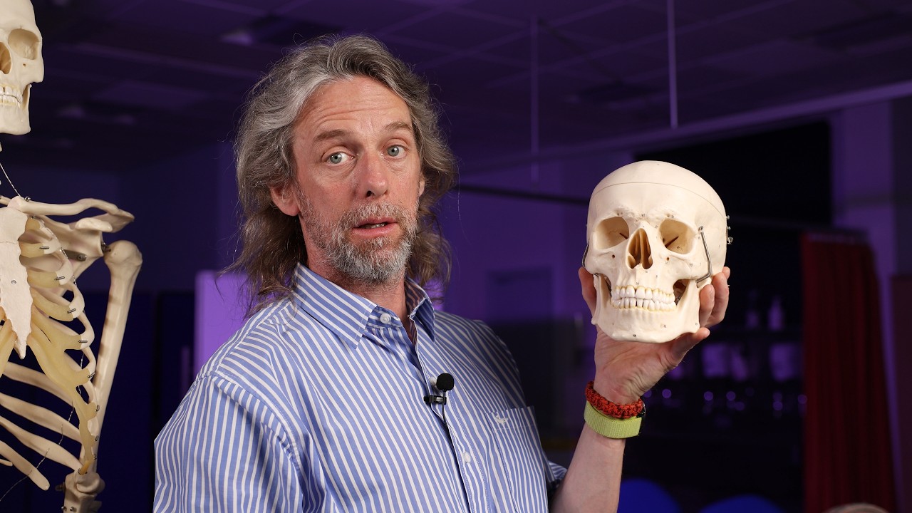
Neuroanatomy of smell (olfaction)
SamWebster
644 views•2026-05-28

women never forget when you upset them
healsick
745 views•2026-06-01

Your Brain Is Actively Deleting Your Childhood Memories! 🧠🗑️ #Shorts #Anatomy #DidYouKnow
voiceless2345
225 views•2026-06-01

What are you looking at
SuperStaticPro
1K views•2026-05-31
Why Trauma Doesn’t Just 'Go Away'
historyofsimplethings
1K views•2026-05-28


