Dr. Monga masterfully bridges complex neuroanatomy with practical clinical localization, turning dense theory into an indispensable diagnostic roadmap. This lecture is a model of academic precision that provides immediate value for both students and seasoned practitioners.
Install our extension to search inside any video instantly.
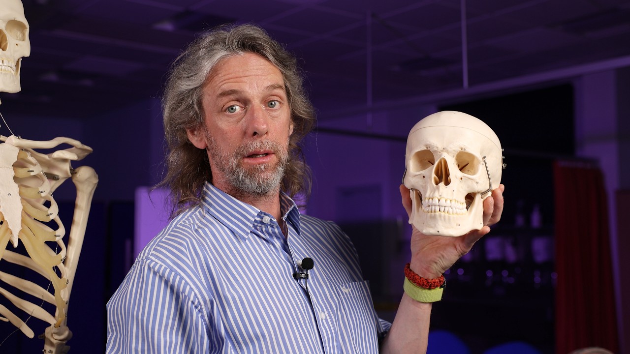
Visual Pathways Visualogy by Dr. Sumit Monga, May 22nd, 8:00 PM ISTAdded:
We live please.
>> Good evening everybody. I welcome you all to yet another I focus online session. Today is lecture number 606. Uh currently we are running in the physiology module number 16. Uh today we have a senior consultant of centersite eye hospital New Delhi Dr. Sumit Mongaser to teach us about the physiology of visual pathway. Welcome sir.
>> Thank you.
>> To moderate today's session we have Dr. Ajinka Viveraesh Muk. Sir has finished his MBBS, DNB and FICO uh in neuroththalmology followed by pediatric ofthalmology and squint uh after which he did his FICO. Sir has done his long-term fellowship at LB Prasad Eye Institute and Arbindi Hospital. Sir is a double board certified of themalmologist specialized in pediatric ofthalmology, stabbismas, neuroopthalmology and cataract surgery. Currently he's a he's a consultant of themalmologist at the I foundation I hospital Bengaluru. Uh sir is a winner of multiple national awards few of which are young researcher award in AIOS 2020 and international athalmic hero award in 20 uh 2021.
Uh sir is a principal investigator and co-investigator in research projects on myopia control OCD angography and optic neuritis. Sir has authored several book chapters and peer at peer-reviewed publications.
Sir has been invited a speaker and presenter at major national and international conferences including AIOC, APA, WSPOS, APGC and INOS. We welcome you sir and over to you for the introduction of our chairperson and speakers.
>> Thank you Dr. Pritika. Thank you for this kind introduction and uh I thank center for site and uh Dr. SH and I focus online for this opportunity. It's been a wonderful platform and a great learning resource. So it's my privilege and honor to introduce today's chairperson Dr. Monalysa Mukatra. Uh ma'am has done her MBBS and MS ofthology from SCB medical college university and she is fellowship trained in pediatricianismas at Arvindai Hospital. She's currently a senior consultant at CFS group at Bhuneshwar and she was a former faculty at Alip Prasad at MTC campus. She has won several best paper uh awards. Uh best paper in pediatricthmology at in Chhattisgarh of society conference 22.
Also the best paper in catact in 23 and best paper in pediatric of themology and Odisa of themological society conference held at held in 2024 and she has se several presentations in various national and international conferences and she has published in peer-reviewed index journals.
Uh I welcome Dr. Sumit Monga who is the speaker for today's uh lecture. uh sir is residency training he has done his residency training at MAMC Delhi and he has completed his F FRCS uh uh at Glasgow and his clinical fellowship trained at Hyderabad and currently working as a senior consultant in pediatric opthalmologist and neuropthmology at CFS group of hospitals in Delhi NCAR he has a vast experience of over 23 years and he has received uh professor bra praash award for best traismas paper at a 2012 12 and he has been awarded with AC Agarwal trophy for best paper in DOSS conference in 2017 and he has received Dr. HShan trophy for best case presentation in 2016 and he has authored multiple book chapters and peer review publications he was a chairperson for a couple of weeks back the session on uh pupil physiology uh and we look forward to learn from the visual pathway physiology from you sir over to you Dr. Sil >> thank you so much uh uh Dr. Dr. Rajen and uh many thanks for this kind opportunity to team I focus Dr. Monica Dr. Monissa my kind regards uh to you as well as Dr. Kitika also and uh warm regards to all the audience which be seeing this at different uh >> all good sir all good sir >> okay thank you so much and obviously uh many thanks to the audiovisisual team for arranging you know the small uh nuggets of the so basically you know I was just going through you know this topic so let me just confide you know that this has been a real personal learning also for us because sometimes when we just venture out in the subject we just tend to forget the basics. So I'm really glad and you know happy that you know uh this was allotted to me because this was a good revision and uh probably a good reinforcement that uh as we go into more complex things uh we should not really uh leave track of the anatomy and physiology because that forms the very basis of you know many of the things. So I'll just try to uh recap some of my experiences you know in of different cases with specific reference to the anatomy of the retino geniculate stride visual pathway which basically means that the visual pathway you know runs from the retina to the uh little genocid body to the stride cortex or the visual cortex and that's what you know we'll just try to uh complete this journey and I'm just uh hoping that you know in the anatomy module a lot of things would have been covered. So I'll just try to just uh recap only the relevant details with more uh emphasis on the physiology. Uh as we move along we'll also uh see how this knowledge helps us to localize the visual field effects and particular attention uh is focused on the cerebral integration of vision. what happens you know once uh when the signals are passed from the eye to the brain and then just a word about you know how does the exonal conduction the optic nerve injury and the regeneration.
So as we all know that basically the route to the vision is a complex one uh starting from the retina to the lateral genic body uh you know to the primary visual cortex and then the path doesn't end in fact uh we have to integrate different parts of you know the uh cortex before we get to see you know uh the image of uh the object in front of us. So as you all know that you know we have these 10 written layers and basically the initiation is when the light is really passing from the eye uh the front part of the eye to the back side of the eye to the retina and simply speaking the light is basically traversing all the layers and going to the photo receptors the rods and the cones and by generation of some action potentials there is a signal signal transaction which passes to uh different layers of the retina. The horizontal sense, the bipolar cells and the amocrine cells which basically comprise the first order uh neurons and then there is a synapse uh between the bipolar cells and the retinal ganglion cells which basically transfer uh the signals from outside the eye. So you can say that retinal gangon cell is the output you know of uh this thing and the um you know the initial uh signal or transaction actually you know passes from uh them from to the outside of the eye. So it's noteworthy to note that you know like normally the cones like a single cone may just uh synapse with a single ganglion cell but there are many rods actually which are uh traversing and synapsing with one ganglion cell and that's probably because uh you know when you have a dark environment probably multiple rods you know when they come into play from passing a signal They help in amplify a dim light source and still gives you some kind of sensitivity although the acquity would be much lesser.
So uh it's a wonderful system u you know beauty that has been set you know for everything. So when the light is good enough of course you know uh you just need probably a single cell u you know to synapse with the younglon cell single cell of cone and that can actually at a very low sensitivity also give you very high acquity. So that's the beauty of you know the initial thing and of course uh you know as the uh conduction takes place in the retina just a pertinent uh point that you know we have a glutamate uh which acts as a inhibitor rather that means uh when you have you know a when you have you know a dark surrounding there are release of glutamate which basically inhibits It's uh the signal and that's what probably the acquity the visual acquity is much lesser you know when you are in a dark environment and when you have conversely you know a lighted environment then you the release of glutamate is minimized and there is no more inhibition of the transmission and the bipolar cells can now release the neurotransmitters toward the ganglion cell that is uh the first sort of neuron to the second or neuron there is a conduction. So this is the role of glutamate and we'll just come back to this why you know I talked about the glutamate a little later. So simply speaking uh we have this arrangement of the nerve fibers which is a very very uh delicate arrangement. We have the horizontal rafe and there are some fibers from the superior and the inferior retina which are being separated.
And uh whenever you have the uh optic nerve uh fibers injury at the level of the retina or say at the level of the optic nerve head. Generally uh we call these kind of defects the visual field defects that we get you know as the nerve fiber bundle defects because these are groups of uh nerve fiber bundles which run in a particular array in a particular organization so that you know we can just get uh what we need. So uh if you see this field effect basically what we are seeing is that uh the right and the left eye of course with the blind spot uh you identify which is the left eye which is on your left and the screen and the right eye on the right of your screen that's how we place the neuropathmic fields. So if you see that uh what we are trying to uh show here is that almost from the disk you know we have these uh aruate shaped defects uh which are happening in the superior fields of both the eyes possibly because of the u uh field effects that you get in glaucoma. So that these superior and the inferior uh fibers the temporal fibers they involve the poles of the optic nerve and because they have a large trajectory uh inside the uh retina that's how you get u this kind of injury to the fibers you know when the IOP is raised similarly if you have an eskeemic damage because of the typical uh blood supply that you have to the retinal fibers we have this classical altitudinal field effect you know of the left eye which means the inferior field of the eye uh you know may be involved.
You can still have a very decent vision 66 69 because even half of the phobia which is spared can give rise to a good vision uh at least in the initial phase and of course you know as the atrophy spreads you have more decrease in the acquity. So another form of number nerve fiber bundle defect is that you know when you have the centroskeal scotoomas the papillomacular bundle which gets involved maybe because of toxicity or you know any other reason majority of cases are you know of toxicity involve the papacular bundle because uh you have the high uh energy taking papomacular bundle fibers which have high mitochondri al demand and things like vitamin B12 deficiency, nutritional deficiency or ethamitol tossy gives rise to these defects and then of course you have the enlargement of the blind spots.
So what we are trying to say is that you know the pattern of the field effects is an important form that where exactly the injury uh you know is in the visual pathway and for the nerve fiber bundle defects at the level of retina or the optic nerve head these are the common field effects that we see. Now as we just go along from the eye uh we uh you know the pre-casmal uh quadrant and if you see uh the in the cartoon if we see this u involvement so suppose you know we have the optic nerve with the precasmal uh junction if it's involved so maybe one half of the complete left eye can be involved which is shown by a complete scootoma in the left eye and uh in the right eye because there are some fibers which are crossing uh the the nasal fibers crossing the nasal uh retinal fibers which are crossing at the level of kasm they're looping uh in the form of a wibbrand's knee when that gets affected you can have a supra temporal field effect a very localized field effect that you to really look forward you know and then uh god this kind of thing. So this is the classical uh junctional scotoma uh which is formed by a precasmal lesion. An important thing to note and distinguish is that you can actually have a monocular field effect. Note that junctional scuttoma was basically a bilateral uh field effect where you have involvement complete involvement of one of the optic nerves and the one beliban fibers the looping that is happening uh that is giving rise to a superbral feed in the other eye also. So it's a bilateral field effect uh the junctional scoma. While if you have a junctional compression of only say the nasal fibers of one of the eyes uh due to a very localized lesion then you can actually have what you call as a junctional scum of tracheure which is basically an epsilateral field effect because the nasal fibers are gone and that to the inferior mostly this is a super temporal field effect only in one of the eyes.
the other eye may be absolutely normal because uh you may not really get uh any field effect you know there. So this was one of the things and then of course you know if you have the fibers which are getting crossed at the level of the optic chasm and majority of being these the crossing nasal fibers uh say from a pituitary lesion or any their any other cellar lesion you may obviously get a bite heopia which may be complete or incomplete depending on whether it's a inferior compression or superior compression and then you know uh consequently you get a complete uh hetronimmous uh hemianopia.
So this was about you know the pre-casmal or kasmal things. Then as we go back from the kayazma basically what we are saying is that you know after the decision of the fibers which are occurring in the kayazma we have an optic tract which basically has fibers from both eyes. The temporal uncrossed fibers from one of the eye and the cross fibers from the other eye the contental eye. And because we have a very uh large representation uh from the nasal half of the retina to because it projects to the temporal field temporal fields is always stronger. So just imagine that if you have a injury to one of the optic nerves, you would have a dominant nasal deciting fiber of the contental eye and temporal fibers of the ipsil eye. Now because this involves uh one half of the field that means in this kind of right optic tract lesion it would subverse the left sided field. So you will have a left homonymous hemianopia. It may be in congruous uh because again you know as we mentioned that the temporal field effect is much more compared to the nasal field effect.
uh so it may be in congruous uh uh you know and we may also have a contraal RAPD why because you know the crossing nasal fibers they are forming a major chunk of the retina uh of the other eye so you can have a quantal in this kind of thing and one of the other things that we have to uh understand is that in optic nerve tract lesion uh whenever we actually talk about the visual field the center of the visual field is not the optic disc but it is the phobia. So you can just well imagine that you know if you have a optic nerve tract lesion you would actually have uh the involvement of the fibers in such a way that the nasal the fibers nasal to the phobia that means the papular mac bundle uh would be involved in both the eyes. So you may actually have a boat eye atrophy in the other eye and because there is a temporal because there is a uh the polar fibers you know at the top of the optic disc that is occurring you may actually have temporal fiber atrophy that is uh hourglass kind of uh fiber atrophy in the epsilateral eye. So these are all the clues exactly I just wanted to just touch upon them and we just discussed that nautic tract lesion also gives light to a contraal RPD.
So here the journey becomes a little more interesting because now we have to actually go to a region where these fibers which have the second order neurons that have originated in the uh retina. They are now after partial deassision of the fibers are reaching something called a lateral geniculate nucleus or body which is essentially a a lateral part of the phalamus.
So this is a very interesting uh thing because uh when you actually have these visual fibers it's not just going to the lateral genical body. In fact you know there are many parts of these fibers although only a minor chunk is also going to different parts of the brain.
So we have the supraismatic nucleus of hypothalammus which also because the visual input is governing the circadium rhythm also. Then you have some fibers going in the uh controlling the pupilary reflexes as we saw that from the optic tract there is a uh pupilary uh digression that is happening then you actually are having fibers going to the superior colliculus because whenever you see you also your ocular movements also get influenced. So this is important to remember that all the part of the visual pathway is not going just to the lateral genate body but just before the lateral genicate body you have some of the inputs although these are minor part and not really help in the vision but they are actually going to different parts of the brain as well. Now lateral genicate body is one of the very very important uh structure and I'll just like to dwell that in a little detail.
So we all know that uh you know with lot of research in this uh area the neurohysiology we have this uh two set of neurons the magnosellular parroar and in fact uh a minority of the neurons are something you know which we call as a k cells or the conioellular you know which are intermediate between the parosellular and the uh the magnosellar areas. So in fact the layer 1 and two of the lateral genocid body uh receive the input from the magnosellular from both the ipsilateral fibers as well as from the crossing fibers of the other eye from the contal eye. So this is very important to note that the layer one and two uh you know of the little genocid body there are six laminar layers. So one and two are actually you know getting input from the magnosellar layers and uh three onwards 3 to six are getting signal from the parosellar and these are both ipsilateral inputs as well as the contal inputs. So why this demarcation is important is that the magnosellar neurons basically have very large cell bodies compared to the uh the the cell the soma I mean the cell body is you know big enough that's why they are called magno meaning large and basically these fibers they respond to objects in motion simply speaking you know obviously uh you have other things that you know the gross depth and the small differences in brightness. They're also controlled by the magnosellular uh neurons. While the parocellular or the P cells are basically neurons which have cell smaller cell bodies and which are projecting to layer 3 to six of the uh lateral genate body both from the ipsil as well as from the contental uh eye and there are fibers basically which are ultimately involved in color perception texture shape and fine depth.
So there is a demarcation you know of these fibers which are basically distin to uh probably serve different functions carry different kind of inputs to the brain.
So lateral genicate body why is it important is that it is essentially not only a organ which is relaying you know from second order neuron to a third order neuron. As we discussed that the retinal ganglion cell when initially it synapses with the bipolar cells that's how the disjunction between first and second order neuron is there and the retinal ganglion cell when they synapse in the uh lateral genocid body that's how the second to third neuron happens.
So these are relayed back from the lateral genocid body to the visual cortex. But more importantly the lateral genocid body also receives some selective information from the various parts of the cortex. So why is it important is that when we see around there are lot of signals you know from all over but the brain tells you where exactly to focus. So it has some kind of selective inhibitory signals which helps you to probably disregard some of the things which are not not important and these this is something you know which leads to the gating function of the lateral genocid body. So many of the visual information which may not be very important possibly would not be really transmitted that well here on. So that is why this kind of uh gating is very important to really focus on certain things and all. Of course there are other aspects of uh perceiving a image.
We'll just talk about it when we talk about the uh posterior visual pathway.
So from the lateral genicate body if you see it's a multilamer structure which are supplied which is which is having uh multiple uh kind of arterial supply. the blood supply. So that makes it very interesting that suppose anybody gets although LGB lesions are very rare but suppose someone gets a uh lateral genocid body lesion here you would actually get something called a wedge-shaped defect or centralopas that means you would have a very sector of involvement which may be homonymous again that is one side of a field will be involved depending on which right or left gen is involved So you have essentially two arteries supplying the central part of the u lateral genetic body or the hilum that is supplied by the posterior coridal artery while the lateral and the middle horns are supplied by the anterior coridal artery and depending on what kind of defect it is what kind of uh eskeemic damage you know to the lat licen body that happens you can have a various uh very distinct sectors you know which will be involved depending on you know whether it's a posterior cororoidal artery lesion or a anterior coridal artery lesion and then you can actually have vertical sectors also involved which can happen because of bilateral LGB lesions. So one needs to be you know aware of these kind of sectors uh getting involved.
So from the lateral genocid body the fibers the optic radiations to be precise they pass u initially fanning out and then probably consolidating you know to both the occipital loes. What is interesting to note is that the fibers from the uh superior u the super from the superior half of the lateral genicate body which are essentially the fibers you know supplying the superior retina which is controlling the inferior field. They have a little uh kind of a straighter course from lateral genetic body to the uh occipital cortex. While the inferior fibers because they come in contact with uh the u the lateral ventricle they have to just uh kind of groove you know over the uh the temporal horn of the lateral ventricle and that is why they actually form a loop in the temporal lobe uh which we all know as the mayor's loop because of this uh anatomical you know kind of detour that they have to take you know from the routine uh the mayor's loop really you know forms a very important uh distinguished uh you know because if this part of the uh radiation the optic nerve optic radiation is involved then it classically you know gives to gives rise to a very classical field effect we'll just see see in a while so basically uh from the uh lateral genicate body these radiations are going to what we call as the primary visual cortex.
Uh there are similar names that have been given to this area. If you uh just check it out. Uh some people call it the area V1. Uh there it was also called the broadman area 17 and it is also called the striat cortex. So the very interesting uh thing which I came actually I in fact uh just uh went to you know the Adler's physiology that we had studied you know in our MBBS and very interestingly you know I just uh kind of uh stumbled upon one of the fact that initially you know when the visual cortex was uh found you know there was this u young scientist called Jinary you know he just was seeing you know some of the uh brains from you know the cat and he just stumbled on you know a very distinct path which appeared like a strike you know on the back side of the brain he never knew that you know what exactly its function was but then you know if you remember from our neuro anatomy there is something called a stry of generary so it's basically you know uh named after him and these are quite distinct fibers that to actually uh uh you know can see in a cut section of the brain. Of course you know after that you know a lot of water has flown under the bridge and the researchers came across you know the visual cortex. I mean finally it turned out to be visual cortex and basically you know if you see uh in the adjacent diagram here. So when we say area 17 the broadband area 17 we have other areas uh both in the motor cortex region the prefrontal cortex region all over the brain uh and the visual areas you know which subverse the vision are mainly the 17 18 and 19.
Actually the main vision is formed you know in the area 17 the visual cortex and the uh secondary cortex uh sometimes we call you know as the uh adjacent area which is the 18 and uh the 19. So these are also part of the visual association that means you know there are some fibers coming from the primary visual cortex which go to these areas as they go to other parts of the uh cerebral cortex and as the vision is integrated.
So primary visual cortex basically has a laminar organization which uh really helps it pack a lot of things because there is a very complex uh circuitry that is involved you know in forming the vision. So simply speaking we have these six layers you know of the primary visual cortex. Essentially the layer one is not actually a layer. It is basically involves the dendrites but just for the names sake you know we take it as a number one layer. The other layer of course you know have uh you know the different exxons which are running through it.
uh if I may just draw your attention uh it's basically the layer four of the visual cortex you know which you can say is the main input layer you know whatever the fibers that are coming from the lateral genocid body uh are mainly going to layer four of the uh primary visual cortex. The other layers are essentially you know the uh areas which are relaying the information from this part to the other parts or from you know one part of the visual cortex to the other. So that's why we see that you know the pyramidal cells which are very large cells you know which are traversing the different uh layers of the visual cortex and uh when you have these uh layer four of the primary visual cortex is essentially very small concentrated uh spiny shellate neurons that are there. Now if I may just uh uh you know with you recall that when we had talked about the lateral genkin body we had mentioned that you know both the M and the P that is the magnoselar and the parocellular neurons you know as they're coming from the retina because these are essentially retinal gang cells they are basically basically you know to the natural genocid body of one side helping you get the inputs both from the ipsilateral site as well as the contrlateral side. So when you actually have these fibers synapsing in the lateral genate body, the contraateral neurons, you know, from the other eye, they're essentially synapsing in one layer 1, four and six.
And the ipsilateral side neurons are essentially which are basically formed because of the temporal fibers you know the uncrossed fibers that are coming to the laterant body are essentially you know synapsing in layer 2 3 and five you know so just have a rough idea I'm just going in sequence so the correlate is that when these fibers come from lateral genocin body to the visual cortex And as we just discussed that the layer layer four is the main input layer you know of uh the primary visual cortex you know where the fibers from the lateral genocid body come. So you can see by color coding that the uh magnosellar as well as parosellar neurons are coming to layer four layer four of the lateral genetic body and in that also if you see carefully the green coded magnosellular layers they're projecting to what you call as a layer 4 C alpha these are subunits you know of that layer 4 and the power cellular layers are essentially uh you know coming and giving their inputs to layer 4C beta. So this is uh something which you should know in the background. Now that's a very interesting thing that uh we have to understand that because there is a limited space how the nature does it it just uh has stacked you know this information in the form of a cell columns. So in the primary visual cortex cells uh you know they are arranged in layers and the layers are arranged by their preferred orientation meaning thereby that these specific cells which are probably having a similar orientation. They are orderly arranged in the form of columns and in the columns also because we have input from both a monocular or ipsilateral side and we have input from the other side also you have stacking of you know intermittent stacking of you know uh things from either one eye or sometimes both eyes. So that is a concept that is doing the rounds you know in uh lately for the past decade and that gives rise to the concept of ocular dominance columns. So basically what are ocular dominance columns?
They're alternating stripes of neurons in the primary visual cortex that respond preferentially to input from either one of the eye that is the right or the left or from both eyes. So in fact uh what is happening is that the uh uh the researchers have found that there is an increasing evidence that you know in the stride cortex you have ephrine fibers uh which are coming uh which are connecting the bilateral binocular cortical cells that respond to stimulation of either the one of the eyes or you have monocular cortical cells that respond to stimulation of only one eye. In fact, humans, you know, they have seen that 70% of the cells in the stride cortex are binocular while monocular are say minority 20 to 30%. So this is huge significance because if you may have actually related it that lately we have something called a doptic therapy for the amilopia management. So what it means is that even if you have a unilateral deprivation of vision due to one or the other cause leading to amopia you know in the childhood in the critical age sometimes stimulation of these binocular cortical cells by a binocular stimulus which is essentially the uh concept of dioptic therapy. You may actually get over that amlopia. Of course, you know, this trimlessness has to be in such a way that it is providing more clearer image to the dominant eye and the lesser sorry to the amlapic eye, beg you pardon. And the uh better eye, you know, has a little lesser uh intensity stimulus, but both eyes are open. So, you're not really theoretically patching. So this is a very very important concept you know and a major transition you know from the concept that a uniquaria just needs uh you know kind of a penalization of the better eye. So because of the binocular cortical cells the representation is binocular and because we have cells you know which have essentially inputs from both sides the right and the left still a unateral amlapia can have treatment uh with stimulation of bilateral you know binocular stimulation.
So this is the concept you know that I wanted to just put across. So we also know that you know there is a specific arrangement of fibers you know as we go from the retina to the optic nerve to the optic tract. So briefly if you see the macular fibers just see in the optic nerve head junction you just see that it's essentially a temporal part of the optic nerve. So by the time it reaches the entropical nerve it really confi uh confines it to the central area and in the kayazma in fact it's towards the posterior part of the kayasma that you know these macular fibers in there in the optic nerve tract then again there is a little rearrangement of fibers and that carries on from the little to the little genuine body and basically what we are uh you know having is that whatever arrangement that we had. If two cells have been arranged, you know, in a particular order in the retina, the same order gets transmitted all throughout the course of the visual pathway to the visual cortex.
And this is important because the spacing the spatial localization of the images would be possible because if whatever uh you know spatial difference that you have in the retina that doesn't correspond to the visual cortex then of course you know you would have a different spacing when you actually see that particular thing and then of course you know uh one has to understand that there is a lot of overlapping visual field you know when you see with both eyes. So these are not mutually exclusive and the lower cartoon you know will help you understand that you know when you have the phobia the macula that is represented although it's just a pit a small area in the retina but it has a large representation in the visual cortex the area 17 you know in fact majority of that the posterior pole the occipital pole for that matter is contributed you know by the color coding is helping you understand that's the phobia And then the peripheral part of the retina is much less area coded you know in the visual cortex. And because this small phobia you know which is really uh you know seeing that image uh and that image has a bigger representation in the uh occipital cortex that accounts for something called cortical magnification.
Otherwise you would have actually you know because as you know that you know when the uh image is transmitted through the cornea the lens if you remember the physics obviously the the inversion of the image takes place and then you know it's a minified image but by the time it reaches the visual cortex it's again magnified you know because of these principles. Now very important uh this thing to understand that suppose if I ask you to just see a image you know on the right side if you would see that what you seeing you are just seeing small dots black and white dots believe it or not when the image reaches the cortex it is just this and when the fibers you know from the occipital cortex the dorsal stream what we call as a dorsal stream go from the occipital cortex to the parietal lobe.
It helps you understand and map that where do we have to concentrate. So probably out of those you know like blobs of the the black and white images it is helping you focus that you know look probably this is the demarcation that we have to see. Now when you actually have inputs from the occipital cortex the same image will also go to something called a ventral stream to the temporal loes. Now it will it is the Google of our brain. It will help you understand that look I'm concentrating on this image now possibly it's a dog it's an animal I have seen it earlier.
Okay. So this is what the occipital cortex has done is it has just uh you know seen the image. What the parietal cortex or the v dorsal stream has done is it is probably helped you focus that where exactly you have to concentrate and what exactly it is has been found out by something called a what system or the vententral stream and how to react to that image. Is it something dangerous that you have to really go or we have to go close to that uh particular creature and cuddle it?
The frontal lo type. So basically if we are actually you know seeing the front part of the uh brain is helping us think.
Middle part of the brain the parietal and the temporal lobe is helping us map and do that's something called a where and what pathway. And obviously the images were formed in the occipital cortex. So this is the cereble ination that is very important that the occipital cortex is not really helping you recollect what it is. And there comes a concept that you know when you have the affliction of different parts of the brain uh how it fares up. So if you have just the occipital radiation or the oxipital lobe we have something called a cerebral visual impairment. So if the vententral part the parietal loes you know and all those things are also involved along with the central area of the brain which is basically the middle brain uh the uh brain stem you can have cerebal pulsy along with CVI and if it's a large involvement even the person cannot think the complete brain you know is eskeemic we have multiple disability where you cannot comprehend also obviously you have ver visual impairment so you have something all agnosas and neglects. Basically what he's saying is that you know this comprehension that where exactly what exactly is the thing.
So if you have some lesions of parietal and the temporal loes and the where and what pathway is affected you will lead to something you know which is called agnosia unable to comprehend. So if you are not unable to perceive what it is that's the vententral pathway we just found that what exactly it is patient is unable to recognize you know like what exactly uh you know is what he's seeing if you are unable to associate because remember the temporal lobe was the Google lab you know of our brain so we have seen it we have registered it that this is what it is supposed to be so if you see an animal probably you know you would uh understand it's a carnivore herbivore or it's a pet you know and this is something you know which temporal lobe or the vententral pathway does. So if you are unable to recognize your new relatives or any celebrity possibly that is again because the inability to recognize that's called proopagnosia that means you are unable to identify the faces you know again a temporal lobe lesion balance syndrome the dorsal or the wear pathway so even if you know you are able to uh see a particular thing you are unable to you know really relate you know where exactly you have to focus So a balance syndrome specifically you know leads to this. So if you just quickly see basically you can have different afflictions uh when you have CVI patients which is not which is not unusual in our practice they see better in familiar environment.
Uh clutter makes the vision difficult.
They have a strong preference for a red and yellow because again you know the color association areas are the uh are little away from the visual primary visual cortex and mostly they have a inferior field effect that's because of the uh the parietal lobe lesions or the eskeeia they are unable to recognize the figures in the faces. So if you have a very severe def deficit you know of CVI the several visual impairment patient is not able to fix that's called a low functioning CVI and a high functioning CVI is when you are just having a processing difficulty of complex visual information but still you are able to you know see but you're not able to recognize you're not able to recognize and process the speed. So all these lesions in the parietital or the wherein and the what pathway will lead to deficits in motion perception which is called ainato topsia or deficits in color vision which are little higher order functions. So these retrocasmal lesions uh specifically of the occipital lobe they can either uh lead to quadrantic field effects if they involve the optic radiations like a parietal lobe lesion would lead to probably something called a pie in the floor uh inferior quadrantopia or a temporal lesion would be pi in the sky. And then of course uh if you talk about the occipital lesions we just discussed that actually uh it can lead to a very congruous uh lesion with or without macular sparing.
So why macular sparing? Because if the occipital pole is paired because it has a dual blood supply it would probably lead to u sparing because of the dual blood supply from middle cerebral and the uh the posterior cerebral artery.
the lesion would have a macular sparring. And then very interestingly, if there is a specific occipital tip lesion, you can actually still have a homonymous uh kind of a central scotoma uh which would be you know just because you know the occipital lobe the macular representation area is involved in both the eyes. Then just a quick word about you know the exonal conduction. So you know that there are action potentials you know which transmit these signals and luckily you know we have this myelin you know as the optic nerve goes from the optic nerve to different parts of the visual pathway. The myelin basically decreases the capacitance and less uh sodium gated channels are needed to depolarize the exxons and it increases you know the passage the ease of passage the fastness uh the swiftness of the signals and of course you know when you have a demalating lesion you know of the exxones uh you have a conduction deficit as happens in the optic neuritis and other demalating lesions. So this can be akin you know to a dis a wire with a deteriorated sheath.
So of course you know the conduction would get delayed or sometimes get affected in this. So exonal transport in a nerve basically you have the slow exonal transport or a fast exonal transport u and when you know we have the orthog transport again uh that is going from cell body towards the brain from you know closer to the eye towards the brain and then you have some retrograde exonal transport from the brain towards the eye. So how do you really have a optic nerve injury or a vjury to the visual pathway? So if you have blockage of external transport of any kind or you have demination like in optic neuritis or sometimes you know at the level of the retina because some reason there is an excess glutamate you know we talked about it you know that when excess glutamate is there it inhibits transmission you know in the retinal area and then of course you know certain kind of damages can also lead to free radical uh formation. So all these are you know pathologies which lead to the exxon damage and if at all the exxon repair you know has to happen you know in the visual pathway uh as we you know like suppose we are treating the optic neuritis what is exactly happening is that we are actually promoting remalination you know using that imosuppressive therapy the steroids and the other things or you can actually also do it by neurop protection that is blocking the exit toxins ity or administrating the neurotrophic factors, inhibition of apoptosis at various stages. These are all research uh you know uh food for these are all things that you know uh gives you food for thought that in coming times how do we will manage you know the optic nerve lesions of the uh visual pathway. So just to summarize just to leave the last 10 minutes for discussion. So visual pathway obviously is a complex signal interaction between the retina and the lateral genocid body and occipital visual cortex and the visual association areas. There is a complex cerebral integration of vision and it's not just that the visual cortex is responsible for vision alone. We have the other loes which are also contributing. The visual pathway disruptions in general can be localized by characteristic uh visual field effects and the visual pathway nerve injury can occur either by demination, exonal transport disruption, glutamate toxicity or free radical formation and exoral uh regeneration is difficult but uh possible. Thank you so much.
Thank you Dr. Sumit was very extensively and very nicely covered. It's such a complex topic but you really made it very simple and uh demonstrated the clinical relevance of all those uh regions and areas getting affected. I'm sure that this will serve as a very great tool for all the postgraduate and fellows out there to know the uh basic physiology of the visual pathway. Uh Dr. Monalysa, do you want to add any comments from your side?
>> Uh thank you Dr. Sumit. Uh very wonderfully Dr. Sumit presented about the uh physiology of visual pathway. Not only physiology, he also covered the anatomical part. He also covered the higher visual function areas and demalanetic le and everything. Uh so just quickly I would like to summarize that the optical system of eye mainly the cora and lens uh they act like convex lens. So because of light refraction through corna and lens the image formed on retina it is inverted.
It is inverted vertically and horizontally and also smaller in size than the actual object. So the object in upper visual field it falls on inferior part of the retina. Object in the lower visual field it falls on the uh superior part of the retina. From the right side falls on the left half of the retina.
From the left side it falls on the right half of the retina. So each eye receives uh information from both visual hemifield. So the retinal photo receptor they convert the light into neural signals which then pass through the optic nerve and this inverted retinal image map it is preserved through the optic nerve also. Then the signal goes to the optic where the nasal fibers they cross and temporal fibers they remain uncrossed. So now the left visual field from both eyes it travels through the right optic tract and the right visual field from both the eyes it travel through the left optic tract. Okay. And the same information it uh uh travels through lateral genulate body and optic radiation and finally it reaches the visual cortex. So in the primary visual cortex uh this inverted map is also maintained. uh but we do not see any object uh reversed. Okay. So uh how it happens uh our brain does not flip the image like camera does but we perceive the world erect because the brain uh it learns spatial relationships. So the visual cortex and the associated area it create a meaningful spatial perception.
So thus uh although the retinal image is inverted the brain uh interprets it as upright. Then yes uh from occipital cortex the visual information it goes for further processing through the vententral stream and the dorsal stream and uh if uh this this is affect and this affection we see in cases of CVI patients. So the clinical intercorrelation of uh visual pathway is actually to localize the lesion in uh respect to the visual field. So if we see that one eye field is affected that means other eye field is normal then mostly one eye uniopular optic nerve is affected. So if we see bmporal hemianopia then probably the lesion is at the opticisma level. If there is homonymous hemiopia then mostly it is at the optic tract level and it is mostly in congruous type that means a symmetrical in both uh the field. So periquadronopia already uh everything is discussed by Dr. Sumit just I'm touching it quickly. So periquadronopia uh it when uh the lesion is at the temporal lobe and inferior quadrantonopia when the leion is at the paral lobe. Then occipital cortex leen uh it will give rise one homonymous hemianopia field which is congruous in nature and most of the time sparing the macula because it has the dual uh Black.
>> Thank you Dr. Monison. It was a good overview which you gave and I think uh it was quite interesting to learn about basic physiology and how it works and how it affects the clinical uh scenarios and we can localize by understanding this physiology uh by sitting in the clinic and looking at the patients fields and presentations to us.
Thank you Dr. Pritika. Uh thank you for this wonderful session. Thank you Dr. Sumit and Dr. Monalis again. Over to you Dr. Kitika. You have some announcement to make.
>> Yes, a very very elaborative class sir.
Uh thank you so much for the class. Uh so not to forget for the audience a gentle reminder. Uh we have a physical focus program from July 5th through 12th. It's a national post postgraduate education program in of themology that's going to be held in Hyderabad this year.
Uh which will have both direct session and hands-on sessions. Uh so hurry up.
Registration is limited to 300 300 candidates only. You can register online with the below given link. Uh and uh do not miss the opportunity to sharpen your skills and learn from the future leaders and present leaders in of themology.
>> Thank you.
>> Thank you.
>> Thank you. Thank you everyone. Thank you so much.
>> Thank you sir. Thank you.
>> Thank you everyone. Thank you.
Related Videos

Recovery pronouns. Neuroplasticity & practical neuroscience tips to help recover from pain & fatigue
Fantasticneuroplastic
907 views•2026-05-31

No Eyes, No Darkness? 👀😱
Huwatif
630 views•2026-06-02

I Saw the Thing Crash. Then I Lost Hours | Beyond Black Budget
BeyondBlackBudget
148 views•2026-05-30

Your Brain Is Actively Deleting Your Childhood Memories! 🧠🗑️ #Shorts #Anatomy #DidYouKnow
voiceless2345
225 views•2026-06-01
Neuroanatomy of smell (olfaction)
SamWebster
644 views•2026-05-28

What are you looking at
SuperStaticPro
1K views•2026-05-31

Size Illusion
WTFactt_t
1K views•2026-06-03

Why Trauma Doesn’t Just 'Go Away'
historyofsimplethings
1K views•2026-05-28


