The video masterfully transforms a macabre curiosity into a sophisticated study of molecular resilience, proving that even our most fragile organ can achieve a grim form of immortality. It is a rare example of science communication that successfully balances sensationalist appeal with genuine academic depth.
安装我们的扩展,即时搜索任意视频内容
These Prehistoric Brains Survived for Millennia and the Explanation Is Genuinely Disturbing本站添加:
Number one. Alexander Morton Heyward's 4,400 preserved human brains. The human brain is one of the wettest organs in the body. It is close to 73% water and much of the rest is fat.
Everything we know about decay says it should be among the first things to go.
Within days of death, the brain's own enzymes begin to digest it from the inside.
Bacteria flood in, the soft tissue turns to liquid and drains away. By the time a body is reduced to a skeleton, the brain should be long gone. And yet again and again, archaeologists open a skull that holds nothing else, no skin, no muscle, no organs, and find a shrunken brain still sitting inside. Alexander Morton Heyward is a molecular paleobiologist at the University of Oxford. In March of 2024, she and her colleagues published a survey that reframed this entire puzzle.
They assembled an archive of 4,405 preserved human brains. The records stretched across more than 200 separate sources and over 300 years of writing.
The brains came from 12 countries and six continents. Some came from Arctic permafrost, some came from desert graves, from shipwrecks, from battlefields, from the cemeteries of old asylums, and from burials thousands of years old.
The oldest brains in the data set are around 12,000 years old, reaching back to the end of the last ice age. The most startling figure in the study is this.
In more than 1,300 of these cases, the brain was the only soft tissue left in the body.
Everything else had skeletonized.
The bones were bare.
But the brain remained.
This directly contradicts the normal order of decomposition, in which the brain is supposed to be among the first organs to disappear, not the last to survive.
Morton Heyward identified five different ways a brain can be preserved. The first is dehydration, where the tissue dries out. The second is freezing. The third is saponification where fat turns to a waxy substance.
The fourth is tanning, the same kind of chemistry that turns hide into leather.
And the fifth is something else, a mechanism that does not match any of the others and that seems to apply specifically to those brain only cases.
Her leading explanation for that fifth pathway is molecular cross-linking. The idea is that inherent proteins inside the brain bond together into a stable, decay-resistant polymer. The brain has a heavy blood supply, so it is rich in iron. Under the right conditions, free iron can drive oxidation reactions that lock proteins and fats together into a solid mass.
The closest everyday comparison is the way oil paint hardens as it dries or the way leather is tanned. It is a reaction that, once started, arrests further decay and converts soft tissue into something durable. The preserved brains shrink often to about a third of their original volume, but they keep their shape. Researchers can still see the folds, the ventricles, the division into hemispheres.
Earlier studies even found intact neurons and blood vessels under the microscope in brains from a 2,600-year-old British burial and from a mass grave dating to the Spanish Civil War. The shared environment in nearly every case is fine-grained waterlogged sediment with very little oxygen, the kind of setting that starves the microbes of what they need to feed.
Morten Haywood describes the texture of these specimens as something like tofu, soft and rubbery, a sign of those polymerized proteins.
The disturbing part of her conclusion is not that brains can survive. It is that the brain may be uniquely predisposed to preserve itself, which means countless examples may already be sitting in graves undiscovered simply because no one expected them.
Excavators often throw them out, mistaking the dark mass for mud or assuming that soft tissue cannot possibly last. If she is right, the most fragile seeming organ we have is quietly one of the most durable.
And if simple chemistry can do this over 12,000 years, the obvious question is what happens when a brain is not slowly buried, but instantly cooked?
Number two, the Herculaneum vitrified glass brain.
In the year 79 of the common era, Mount Vesuvius erupted and destroyed the Roman towns around the Bay of Naples. Pompeii is the famous one, but its neighbor, Herculaneum, was buried in a different and more violent way.
Instead of a slow rain of ash, Herculaneum was hit by pyroclastic surges, fast-moving clouds of superheated gas and debris.
In one building, the Collegium Augustalium, a young man of about 20 was found lying face down on a wooden bed.
He almost certainly died in seconds.
The forensic anthropologist Pier Paolo Petrone at the University of Naples Federico II examined the remains.
Inside the skull, he found something no one had ever documented in human remains before, glossy black fragments like shards of obsidian. He published his findings in the New England Journal of Medicine in January of 2020 and in the journal PLoS ONE later that year.
The fragments were brain tissue that had been vitrified, meaning it had turned to glass.
This is the only known case in the world of human soft tissue converted into glass.
To understand why this matters, you need to understand what vitrification actually requires.
A material becomes glass when it is heated to a high temperature and then cooled extremely fast, so fast that its molecules freeze in place without arranging into crystals.
This is a delicate, specific process. It does not happen by accident very often.
Petrone's team estimated that the temperature at the moment of death exceeded 510 to 520° C.
They worked this out from charred wood near the body, which carries a chemical record of how hot it got.
The sequence seems to have been a brief blast of intensely hot ash followed almost immediately by rapid cooling as that cloud dissipated. That is exactly the recipe for turning tissue into glass. To prove the glass was really brain, the researchers used proteomic analysis, which identifies the specific proteins present. They found proteins known to be expressed in adult human brain tissue. They found fatty acids characteristic of human brain fat and human hair. They even detected the genes that code for those brain proteins. The victim's spinal cord, inside the bones of his back, had also turned to glass.
Under a scanning electron microscope, the team saw preserved network structures that they interpreted as neurons and the fibers connecting them.
In 2025, a follow-up study led by Guido Giordano, published in Scientific Reports, refined the picture.
The argument is that the vitrification was caused by a cloud of superheated ash at over 500° that arrived first and dissipated within moments before the slower, cooler pyroclastic flows buried the town. The man's indoor location was crucial.
Sheltered from the main flows, his body was exposed to that brief extreme pulse of heat, hot enough to vitrify, but gone quickly enough to leave glass rather than ash.
What makes this so striking is that organic vitrification, the conversion of living tissue into glass, had never been observed in nature before this case. We use vitrification deliberately in modern science to preserve cells and embryos by freezing them so fast that ice crystals never form, and even to lock up nuclear waste in stable glass. Here, a volcano did it to a human brain by accident almost 2,000 years ago.
Heat preserved what it should have destroyed.
And if a brain can survive being turned to glass in seconds, it raises a different possibility entirely, that a brain might survive not for thousands of years, but for hundreds of millions hidden inside solid rock. Number three, Coccocephalus wildi, the 319 million-year-old fish brain.
Step back now from human history into deep time.
Coccocephalus wildi was a small fish that lived about 319 million years ago during the Carboniferous period, long before the first dinosaurs. It was a ray-finned fish, part of the enormous group that includes most fish alive today.
Only one fossil of it is known, a single skull recovered from the Mountain Fourfoot Coal Mine near Lancashire in England.
That skull had been sitting in the collection of the Manchester Museum for roughly a century. No one knew it held a brain.
In 2023, a doctoral student named Rodrigo Figueroa, working at the University of Michigan with the museum director Matt Friedman, scanned the skull.
The discovery was published in the journal Nature in February of that year.
The remarkable thing is how it was found.
Figueroa was using computed tomography, a CT scan, to study the inner ear anatomy and the structure of the skull.
The skull had never been broken open.
And inside the intact braincase, the scan revealed a dense three-dimensional object with the unmistakable shape of a brain.
This is the oldest example of a well-preserved vertebrate brain in three dimensions ever found. It has a central body with clear left-right symmetry.
It has hollow spaces that correspond to the ventricles.
It has at least one cranial nerve and other filaments extending outward, exactly where you would expect nerves to leave a brain.
There was a surprise in its anatomy.
In Coccocephalus, the brain folds inward on itself.
This is the pattern seen in living sharks and in other early branching fish.
But in modern ray-finned fish, the brain develops by folding outward, a reversed arrangement.
Because Coccocephalus sits near the base of the ray-finned family tree, its inward-folding brain suggests that the outward-folding plan of today's bony fish evolved later than scientists had assumed.
A single fossilized brain shifted the timeline of a major evolutionary change.
The preservation happened because the fish was buried quickly in an environment with very little oxygen and the right chemistry to mineralize the soft tissue before it could rot.
The brain became denser than the sediment that filled the rest of the skull, which is exactly why it stood out so clearly on the scan.
The fish itself was small, perhaps 15 to 20 cm long.
It probably lived in an estuary or a coastal swamp eating small crustaceans, insects, and squid-like animals.
The deeper implication of this discovery is unsettling in a quiet way.
If a brain can survive 319 million years inside an ordinary-looking fossil skull, and if it can go completely unnoticed for 100 years in a museum drawer, then how many other preserved brains are sitting in collections right now? Never scanned, never suspected.
Figueroa and Friedman argued that brain preservation may be far more common than anyone realized and that it has simply been missed because no one was looking inside the skulls.
And once you start asking how far back the record of fossil brains goes, you arrive at a creature that lived 200 million years before this fish. Number four, Fuxianhuia protensa, the 520-million-year-old arthropod brain.
To find the oldest fossilized brains on Earth, you have to go to the Cambrian period more than half a billion years ago to a place in Yunnan province, China, called the Chengjiang biota.
There, in fine ancient mudstone, lived a creature named Fuxianhuia protensa.
It was a stem arthropod, a distant relative of insects, crustaceans, and spiders with an elongated body and a broad shield over its head. It lived about 520 million years ago.
And it carries one of the oldest fossilized brains and nervous systems ever described. The key work was led by Nicholas Strausfeld, a neurobiologist at the University of Arizona, together with Xiaoya Ma and Gregory Edgecombe. Their landmark paper was published in Nature in October of 2012. In Fuxianhuia, the brain is preserved as a flattened film of carbon, darkened against the pale rock.
When they traced it, they found a three-part brain divided into regions comparable to the brain organization of modern insects and crustaceans. This was a shock.
A three-part structured brain in an animal this ancient implied that complex neural architecture appeared very early in the history of life, far earlier than many had expected.
The preservation is thought to have happened through rapid burial in fine sediment with no oxygen, where the neural tissue was replaced by a carbon film before decay could erase it.
A follow-up study in 2015 described more specimens and reinforced the conclusion that these structures were genuinely brains.
This is where the story becomes a scientific battle, sometimes called the fossil brain wars.
Critics argued that brains decay far too quickly to fossilize at all. They suggested the dark traces might be something else entirely, perhaps films left by decay-causing microbes, or patterns in the sediment that only resembled a nervous system.
To test the idea, other researchers ran experiments in taphonomy, the study of how things decay and fossilize.
They rotted modern animals under controlled conditions to see what survived and what could mimic a brain.
Strausfeld and his colleagues had a powerful counter argument. The same brain structures appeared in the same positions across multiple separate specimens. Random decay or sediment artifacts would not reproduce identical anatomy again and again. They also pointed to preserved optic tracts, the nerve bundles connecting the eyes to the brain, showing an integrated visual and neural system half a billion years ago.
Note that this carbon film preservation is a completely different process from the mineral replacement that preserved the fish brain we just discussed.
There's more than one way to fossilize a mind.
Over time, as nervous systems turned up in other Chengjiang animals, the weight of evidence shifted and the field broadly accepted that under truly exceptional conditions, brains and nerves can fossilize as carbon.
Fuxianhuia is the deepest reach of that record, an actual map of a creature's neural machinery far older than any brain preserved in bone.
And remarkably, a cousin from these same ancient seas had a brain built on a different plan and a head crowned with three eyes.
Number five, Stanleycaris Herpax, the three-eyed radiodont brain.
In the mountains of British Columbia, Canada, lies the Burgess Shale, one of the most important fossil sites in the world for understanding early animal life.
Among its creatures was Stanleycaris Herpax, a radiodont, which means it belonged to the same group as the famous Cambrian predator Anomalocaris.
Stanleycaris lived about 506 million years ago and it has been preserved with its brain and nervous system intact.
The study was led by Joseph Moysiuk and Jean-Bernard Caron, working with the Royal Ontario Museum and the University of Toronto. They published their results in the journal Current Biology in July of 2022.
What set this work apart was the sample size.
The team examined 84 specimens.
With that many fossils, they could reconstruct the soft anatomy with real confidence rather than relying on a single, possibly misleading, example.
Many of the specimens preserved not just the brain and nerves, but the digestive tract as well.
Then came the most arresting detail.
Stanley Caris had three eyes. It had two compound eyes on stalks like many arthropods, but between them sat a single large central eye. This kind of median eye had not been seen before in radiodonts, and its size suggests the animal has sharp vision and hunted by sight.
This was a small, but highly capable predator around 20 cm long including its grasping front appendages feeding on the tiny animals of the Cambrian seas.
The preserved brain told an even bigger story. It was composed of two segments.
The first, the protocerebrum, was connected to the eyes.
The second, the deutocerebrum, was connected to the spiny frontal appendages the animal used to rake in prey.
The Latin name Herpex actually means rake after those appendages.
This two-part brain contrasts with the three-part brain of modern arthropods and of Fuxianhuia.
It captures an earlier stage in the evolution of the arthropod head showing how these segmented bodies and brains were assembled step by step from soft-bodied ancestors.
By mapping each brain segment to the body parts it controlled, the researchers could test long-standing ideas about how arthropod heads evolved.
The neural tissue itself was preserved as reflective films and mineral traces distinct from the gut and the other organs. Again, the product of rapid burial in fine oxygen-poor mud.
The large central eye, vestigial or absent in many living arthropods, turned out to be an important ancestral feature, a reminder that evolution often discards what it once relied on.
Stanley Keris gives us a second deep time fossil brain to set beside Fuxianhuia, but from a different lineage and built to a different plan.
Together they show that complex brains and sophisticated eyes were present at the very dawn of animal life.
So far, every preserved brain we have met belonged to a soft creature buried in mud or a human caught by chemistry.
But the next case asks whether something far larger, an actual dinosaur, could ever leave its brain behind.
Number six, the Iniopterygian mineralized brain.
Before Coccocephalus made headlines in 2023, there was an earlier discovery that first proved a vertebrate brain could fossilize at all.
It came from a strange group of fish called Iniopterigians, ancient relatives of today's chimaeras, the deep-sea ratfish that are cousins of sharks. The fossil in question is about 300 million years old from the Carboniferous period, recovered from sites in Kansas with related material from Oklahoma in the United States. The study was led by Alan Pradel with collaborators including Philippe Janvier, John Maisey, and Paul Tafforeau.
It was published in the Proceedings of the National Academy of Sciences in 2009.
This was the first fossilized vertebrate brain ever confirmed in three dimensions, more than a decade before the fish brain from the Manchester drawer.
To find it, the team used synchrotron X-ray tomography at the European Synchrotron Radiation Facility in Grenoble, France.
A synchrotron produces X-rays of enormous intensity, which can resolve internal detail down to the scale of microns without ever cutting the fossil open.
The brain had been preserved through mineralization. The neural tissue was replaced by calcium phosphate, the mineral apatite, creating a dense structure that the scan could pick out inside the brain case. This is yet another distinct preservation route, different again from the carbon films of the Cambrian arthropods. There was an important surprise.
The preserved brain was small, and it did not fill the brain case.
There was empty space around it. That detail mattered far beyond this one fossil.
For decades, scientists had estimated the brain size of extinct animals from endocasts, which are casts of the cavity where the brain sat. The assumption was that the brain roughly filled that space. The Iniopterygian showed that in some fish at least, the brain can be considerably smaller than its housing.
That means endocasts can overestimate brain size, a correction that rippled out into how researchers interpret many other fossils.
The preservation happened in a phosphate-rich, low-oxygen marine setting that allowed soft tissue to mineralize quickly before it decayed.
The fossil sat inside a nodule, a hard mineral concretion that sealed the remains and protected them from being crushed. The scan revealed the optic lobes, regions resembling the cerebellum, and the roots of cranial nerves, enough to compare the ancient brain directly with those of living fish.
What this discovery really established was a method. Scan a fossil brain case with a high-resolution tomography, and you might find a brain hiding inside.
That template is precisely what later led researchers to look inside the Coccocephalus skull. It proved that phosphatic preservation, already known to capture muscle and gut tissue, could also capture the brain. And it confirmed that a sparse, but genuine fossil record of the vertebrate brain exists, waiting to be read.
These brains were preserved by minerals forming in the sea.
The next one was preserved by something closer to rot in a still and acid pool.
And at first, it looked like nothing more than a brown pebble on a beach.
Number seven.
The Bexhill Brain Pebble Dinosaur Brain.
In 2004, a fossil collector named Jamie Hiscocks was searching the beach near Bexhill in Sussex, England. Among the stones, he picked up a small brown nodule of ironstone most roughly the size of a pebble. It did not look like much, but it would turn out to be one of the rarest fossils ever found. The only piece of a dinosaur's brain ever identified.
The pebble dates to the early Cretaceous, about 133 million years ago from the Wealden deposits of Southern England.
The study identifying it as fossilized brain tissue was led by David Norman of the University of Cambridge and the late Martin Brasier of the University of Oxford with Alex Liu and others.
It was published in 2016 in a special volume of the Geological Society of London dedicated to Brasier's memory after he died in a car accident in 2014.
He had studied the specimen for years.
The dinosaur is thought to have been a large plant eater similar to Iguanodon and ornithopod common in early Cretaceous England. Under a scanning electron microscope, the researchers found mineralized soft tissues they interpreted as the meninges, the tough protective membranes that wrap the brain, along with networks of tiny capillaries.
They also identified structures resembling cortical tissue and strands of collagen. These delicate features had been locked in by iron minerals and phosphates.
The preservation story is the most revealing part. The team proposed that the dinosaur died with its head submerged upside down in a still acidic, low-oxygen bog or swamp pool.
In that chemical environment, the surface of the brain was effectively pickled. Its decay slowed just long enough for minerals to begin replacing the tissue.
The process is very similar to tanning.
Crucially, the brain had been pressed against the inside of the braincase. So, what survived was the actual surface texture of brain tissue, not just a hollow cast of the cavity. That detail carried a wider implication. If the brain was pressed right up against the bone, it suggests that this dinosaur's brain may have filled more of its brain case than scientists typically assume for reptiles, bringing it closer to the condition seen in birds and crocodiles, the dinosaur's living relatives.
As with the Cambrian fossils, some researchers urged caution, pointing out how hard it is to prove that ancient soft tissue is truly brain and not a biofilm, a mat of microbes that can mimic anatomy.
The most important link for our purposes is the chemistry. The acidic, oxygen-poor, iron-rich water that preserved this dinosaur's brain works in fundamentally the same way as the chemistry that preserves human bodies in peat bogs.
The same conditions, applied across more than a hundred million years and to wildly different animals, produce the same impossible result.
An unremarkable pebble held a fragment of a dinosaur's mind, and that same swamp chemistry, applied to a human body, can do something stranger still.
It can turn us, quite literally, into soap.
Number eight.
The saponified corpse wax brains.
There's a process that can transform a buried human body into a waxy, soap-like substance. It is called saponification, and the material it produces is known as adipocere, or more bluntly, grave wax or corpse wax.
It forms when a body decays in conditions that are cool, moist, low in oxygen, and chemically alkaline.
Under those conditions, the body's own fat undergoes a chemical reaction, a combination of hydrolysis and hydrogenation that converts it into saturated fatty acids and their salts.
This is, in the most literal sense, the same reaction used to make soap from animal fat.
The brain is an obvious target for this process because it is roughly 60% fat by dry weight. When a brain saponifies, it can hold its shape for decades or even centuries, encased in this inert waxy material.
The result is grayish-white, sometimes crumbly, sometimes greasy.
The most famous example sits in the Mütter Museum in Philadelphia. She is known as the Soap Lady, a woman who died around the year 1800 whose body largely converted to adipocere.
The Smithsonian holds a counterpart specimen, the Soap Man.
Both were exhumed in Philadelphia in 1875.
Saponification is one of the five preservation mechanisms cataloged in Alexander Morten Haywards 2024 survey, accounting for a distinct subset of the preserved brains.
Adipocere has a useful property for preservation.
It is hydrophobic, meaning it repels water, and it resists bacterial decay.
Once it forms, it seals the tissue inside it from further microbial attack.
It takes months to years to begin forming, and once established, it can persist for a very long time, as long as the surrounding conditions stay stable.
The conditions that favor it are specific. Clay-rich, waterlogged soil, coffins that hold moisture and drain poorly, mass graves where many bodies create a sealed, oxygen-starved environment, cold temperatures slow the growth of decay bacteria and tip the balance toward adipocere rather than ordinary putrefaction, which is why so many documented cases come from cool, damp climates. A saponified brain shrinks and hardens, but it can retain its gross anatomy, including the folds of the surface and the division into hemispheres.
Forensic scientists pay close attention to adipocere because it can preserve wounds, facial features, and internal organs long after normal decomposition would have erased them.
They can analyze it with gas chromatography to confirm its fatty acid composition and to help estimate how long a person has been dead.
The substance was first described scientifically in the 18th century by French chemists who coined the name adipocere from the Latin words for fat and wax.
What makes these specimens so unsettling is that they look like two things at once, like human tissue and like an ordinary household material. The chemistry of soap making normally carried out in a factory happens spontaneously inside a corpse.
In the same family of conditions, cold and wet and airless, that turns body fat into soap can do something equally strange in the peat bogs of northern Europe. There, it does not make soap of a brain. It tans it like leather and it preserves whole faces with their expressions intact.
Number nine.
The bog body brains, Tollund Man and Lindow Man.
Across northern Europe in Denmark, Ireland, Germany and Britain, peat bogs have given up some of the most hauntingly preserved human remains ever found. These are the bog bodies. They were preserved naturally by the acidic, oxygen-poor, cold water of the peat.
Two of them are especially important for understanding how a brain can survive thousands of years.
Tollund Man was discovered in 1950 in the Bølling Skovdal bog near Silkeborg in Denmark.
He dates to roughly the 4th century before the common era, the Iron Age, which makes him about 2,400 years old.
He was so perfectly preserved that the people who found him believed he was a recent murder victim and called the police.
Around his neck was a leather noose.
He'd been hanged, almost certainly as part of a ritual.
His face survived with a calm, contemplative expression as if he were asleep.
Inside his skull, his brain was preserved, shrunken, but with the folds and the cerebellum still recognizable.
Lindow Man, nicknamed Pete Marsh, was found in 1984 in Lindow Moss in Cheshire, England. He dates to around the 1st century of the Common Era, roughly 2,000 years ago.
His death was violent and deliberate, what researchers call a triple death.
He was struck on the head, garroted or strangled, and his throat was cut.
This combination strongly suggests ritual sacrifice. His preserved brain, like Tollund Man's, retained its soft tissue structure after 2,000 years in the bog.
The chemistry behind this preservation is remarkable. As the moss called sphagnum dies and decays, it releases a substance called sphagnan.
Sphagnan tans tissue in much the same way that tanning turns animal hide into leather. It also binds up nitrogen and chelates, or locks away, the metal ions that decay bacteria need to function.
The bacteria are effectively starved and the body is pickled.
On top of that, the bog water is highly acidic, very cold, and almost free of oxygen, all of which halt decomposition.
There is a strange twist here.
That same acidic water often dissolves bone because it leaches out the calcium phosphate that bones are made of. So, in a bog body, the soft tissue and even the brain can survive while the skeleton softens or partly dissolves.
This is the exact reverse of fossil preservation where minerals replace soft tissue and bone endures.
A related British case, the Heslington Brain, found near York and around 2,600 years old, comes from similar waterlogged, oxygen-poor ground and is the best preserved ancient brain ever found in Britain.
The peat tannins stain everything a deep brown, giving bog bodies their iconic leathery appearance.
The preservation is so faithful that researchers can recover stubble, fingerprints, and even the contents of the stomach.
Tollund Man's last meal, a porridge of barley and seeds, was reconstructed from his preserved gut. The same chemistry that saved his brain also saving his final meal. These bogs preserved the brain entirely by accident, but one ancient civilization took the opposite approach. It tried deliberately and systematically to remove the brain and throw it away.
Number 10.
Egyptian excerebration and the brains the embalmers left behind.
The ancient Egyptians are famous for mummification, for preserving the body so that the soul could use it in the afterlife.
But, there was one organ they did not want to keep.
The brain.
The practice of removing it is called excerebration and it was usually done through the nose.
The Greek historian Herodotus, writing in the 5th century before the common era, described how Egyptian embalmers drew the brain out through the nostrils using an iron hook. The reason is a window into how the Egyptians understood the human body.
They believed that thought, memory, and emotion lived in the heart, not the head.
The brain was considered worthless, so they discarded it. The heart, by contrast, was carefully preserved and left in the body because it would be needed for the final judgment when it was weighed against the feather of Ma'at, the goddess of truth and order.
To remove the brain, the embalmers broke through the ethmoid bone, a thin sheet of bone at the top of the nasal cavity, to reach the inside of the skull. In practice, they often did not pull the brain out whole. They would break it up with the hook or liquefy it and let it drain away, then rinse the empty cavity with resins.
Modern studies using CT scanning and endoscopy on mummies, including work by researchers such as Frank Rühli and Andrew Wade, show that excerebration became common during the New Kingdom, from around 1,500 years before the common era.
But, that it was never applied consistently.
This is where the irony emerges.
A significant number of mummies still contain their brains or large parts of them.
Sometimes the removal was incomplete.
Sometimes it was never attempted at all, especially in earlier periods or for people of lower status who could not afford the most elaborate procedures.
In some mummies, resin was poured into the empty skull and hardened into a solid cast. In others, the brain simply dried in place, shrinking against the wall of the skull.
The Egyptian climate did much of the work. The heat and the dryness, combined with natron, a naturally occurring salt of sodium carbonate and bicarbonate, desiccated whatever brain tissue remained.
Natron drew the water out of the body over a period of about 40 days, mummifying the remaining tissues, including any brain the embalmers had missed.
This is preservation by dehydration, one of Morton Hayward's five mechanisms. The tools have survived, too. Long bronze hooks and spatulas.
In one famous case, an excerebration tool was found still lodged inside a mummy's skull, left behind by the embalmer. The technique varied by period, by region, by cost, and by the skill of the individual embalmer.
Whether a particular brain survived was, in effect, a lottery of method and care.
For modern science, these retained brains are valuable. They allow paleopathologists to study ancient disease, age, and sometimes cause of death using CT scans that do not damage the remains.
And there's a deep irony in it. The one culture that tried hardest to destroy the brain still left us thousands of them preserved by accident. 10 very different cases, 10 very different routes, and yet they all arrive at the same impossible result. The obvious question is what, underneath all that variety, they actually share.
Across all 10 of these cases, the brain is fighting the same two enemies.
The first is autolysis, the process by which the body's own enzymes digest its tissues after death.
The second is microbial decay, the work of bacteria and other organisms that consume the body.
To preserve a brain, you have to shut down one or more of the things these processes depend on, which are water, oxygen, and the activity of microbes themselves.
Each case does this in its own way, but the methods fall into a small number of families.
Dehydration, seen in the Egyptian mummies and in desert burials, simply removes the water that decay requires.
Freezing in permafrost halts enzyme and microbial activity altogether by locking everything in ice.
Saponification converts the brain's fat into adipocere, an inert water-repelling wax.
Tanning in the bog bodies and in the dinosaur's acidic swamp pool cross-links and immobilizes the proteins, much as leather is made.
And mineralization in the deep time fossils replaces the tissue with stable inorganic material, whether calcium phosphate, a carbon film, or iron minerals.
Then there is the sixth pathway, the one Morton Hayward calls unknown, the iron and protein cross-linking that seems to explain the human brains that survive when every other soft tissue is gone.
This is the most intriguing of all because it suggests the brain may carry within it the seeds of its own preservation.
The organ is unusually rich in fat and in iron, and that combination, under the right chemistry, may make it uniquely able to convert itself into a durable solid.
Look at the settings, and a pattern emerges. Rapid burial in fine oxygen-free sediment unites the Cambrian arthropods, the radiodont, the Carboniferous fish, and the iniopterygian.
Acidic, oxygen-poor water unites the dinosaur pebble and the bog bodies.
The Herculaneum brain is the great outlier, achieving in a few seconds of extreme heat what slow burial takes thousands or millions of years to accomplish. And the contrast between the bone-dissolving bogs and the bone-preserving mineral settings shows that preservation is not one process but many, sometimes working in opposite directions.
There is a final, almost poetic detail.
The same iron chemistry that may damage living brains through what biologists call oxidative stress could be the very thing that stabilizes them after death.
The element that helps wear the brain out in life may help it endure in death.
Sites of such exceptional preservation are known as Lagerstätten, and they are where most of these deep-time brains cluster.
But knowing why a brain survives is only half the challenge. The other half is proving that the lump you are looking at really is a brain and not a trick of decay.
The central difficulty in this field is simple to state.
Soft tissue, sediments, and the films left behind by microbes can all look alike. A dark mass inside a fossil skull is not automatically a brain.
So, every claim has to survive a barrage of tests, and the history of the field is full of fierce disagreement. The tools have grown steadily more powerful.
Computed tomography, the CT scan that revealed the Cacops brain, images the internal density of a specimen without ever cutting it open.
Synchrotron X-ray tomography used on the Enantiornithes takes this much further, resolving detail down to a few microns using X-rays far more intense than any hospital scanner.
Scanning electron microscopy applied to the dinosaur pebble and the Herculaneum glass reveals structures at the scale of individual cells and blood vessels.
And proteomics, the analysis of surviving proteins, allowed the Herculaneum team to identify molecules specific to brain tissue, the strongest kind of evidence available.
The doubts are just as important as the tools.
The fossil brain wars sparked by the Cambrian arthropod discoveries centered on whether those dark carbon traces were genuinely neural or merely films left by decay bacteria.
To settle such questions, scientists turned to experimental taphonomy, deliberately decaying modern animals to see which structures survive and which artifacts can mimic a brain.
The evidence that usually convinces skeptics is consistency.
When the same structure with bilateral symmetry appears in the same position across many separate specimens, random decay cannot explain it.
A single specimen is rarely enough, precisely because biofilms can imitate anatomy so convincingly.
The governing principle is to demand multiple independent lines of evidence, anatomy, chemistry, position, and repetition, all pointing the same way.
There's also a conceptual trap that the Eniopteryx helped expose.
An endocast, the mineral infill of the brain cavity, is not a brain. It is a cast of the space the brain occupied, and as that fossil showed, the brain can be much smaller than its housing, which means endocasts can mislead.
Studying human remains adds another layer entirely because mummies and bog bodies carry cultural and ethical weight.
Non-destructive imaging is preferred not only for scientific reasons, but out of respect for the dead. The encouraging reality is that these techniques now let museums revisit specimens collected a century or more ago, finding brains in skulls that have sat untouched in drawers, which raises a genuinely open question. If the tools keep improving, how much of the preserved record is still sitting unrecognized waiting to be found?
Alexandra Morton Hayward makes a striking argument. She believes that preserved human brains are being discovered and then discarded on a regular basis simply because the people excavating them are not trained to expect soft tissue inside a skeletonized body.
If that is true, then the 4,400 brains in her archive are not the complete record. They are merely the ones that happened to be noticed and written down.
The deeper record is almost certainly biased. The deep time fossil brains cluster in a handful of exceptional sites, the Lagerstätte in in places like Chengjiang and the Burgess Shale.
Whole continents and whole stretches of time have produced no known fossil brains at all, which is far more likely to reflect where we have looked than where brains were preserved.
There are glaring blanks.
Mammal fossil brains are essentially unknown despite an abundance of fossil mammal skulls.
Bird brains, despite their delicate skulls, are rarely preserved as actual tissue.
The human brain only cases hint at chemistry we do not yet fully understand, the unknown fifth mechanism that Morten Høyer set out to explain.
Closing that gap is one of the central open questions of the field.
And the prize for closing it is large because preserved brains may hold ancient proteins and perhaps even fragments of DNA that could open new windows on the past.
A brain is a biomolecular time capsule.
The practical path forward is straightforward, if not easy.
Routine CT scanning of fossil skulls in collections, more careful excavation of human burials with the expectation that a brain might survive.
Standardized methods for recognizing and recovering neural tissue.
If those practices spread, the number of known preserved brains could grow dramatically both in the archaeological record and in the deep past.
Museum drawers around the world may already hold specimens that no one has thought to scan. There are, as always, competing views.
Some researchers are optimistic that many discoveries lie just ahead, hidden in existing collections. Others are more conservative, arguing that genuinely exceptional preservation will always be rare, and that we should not expect a flood.
A few speculate about preservation pathways not yet identified at all. The ethics of studying human neural remains will shape how much is recovered and how.
But if these brains really are time capsules, the natural next question is what precisely we can read from them.
A preserved brain is far more than a curiosity.
It can carry ancient proteins that reveal how a body once worked, what diseases it suffered, and what stresses it endured. In principle, markers of neurodegenerative disease might be detectable even in very old brains, offering a glimpse of conditions that affected people thousands of years ago.
The bog brains and the cross-linked human brains have already yielded intact neurons visible under the microscope, structures from minds that stopped working millennia in the past.
The Herculaneum brain expanded the boundaries of what is possible.
Even after being heated past 500° and turned to glass, it preserved identifiable proteins. That a molecule could survive such violence reshapes our sense of what extreme conditions destroy and what they can lock in place.
Each of these cases test the outer limits of biomolecular endurance, and the limits keep turning out to be further away than expected.
For evolutionary biology, the deep time brains are precious data points.
Fuchsian Huya and Stanley Cories show that complex segmented brains existed at the dawn of animal life. Coccosteus revised the timeline for when the modern ray-finned fish brain plan evolved, pushing a key change later than assumed.
The Iniopterygian corrected a basic error in how brain size is read from fossil brain cases, a correction that affects the interpretation of countless other specimens.
These are not isolated marvels. They feed directly back into how the whole story of brain evolution is told.
For forensic science, preserved tissue and adipocere help reconstruct how and when a person died, sometimes long after ordinary evidence would have vanished.
Alongside the brains, these cases preserve other intimate details.
Tollund Man's last meal, the triple death of Lindow Man, the embalming choices of Egyptian practitioners.
Each preserved brain comes embedded in a human or biological story, and the brain anchors it. There are cautions, of course.
Ancient biomolecules can be contaminated by modern material, and they degrade over time. So, researchers must work hard to be sure that the signal they detect is genuinely ancient.
How much of the original information truly survives is a matter of ongoing debate.
But, the trajectory is clear.
The brain, long dismissed as the first thing lost after death, is turning out to be a uniquely rich archive of the deep and recent past.
And that leads to one last question, the one that sits underneath all of this, a question about ourselves.
For most of the history of science, the brain was assumed to be the most perishable part of us, the organ that vanished first, leaving only bone behind.
The evidence gathered here points to the opposite conclusion.
Again and again, in wildly different circumstances, the brain proves to be among the most durable tissues a body has. It is the part most likely to be found intact in shape when everything else is gone.
Consider the span.
From an arthropod that lived 520 million years ago to a Roman who died less than 2,000 years ago, the same organ keeps recurring in the record, and it gets there by an astonishing variety of routes.
Heat that turns it to glass, cold that locks it in ice, acid that tans it like leather, minerals that replace it with stone, the body's own fat converted into soap, dry salt that shrinks it to a hard resin-like lump.
10 different paths, one impossible destination.
The truly disturbing thread running through all of it is that none of this requires a miracle.
It is ordinary chemistry operating under particular conditions.
A brain can become glass, carbon, mineral, leather, wax, or resin, and still hold the architecture of what it once was.
The iron and protein cross-linking that Morten Høyer describes suggests that the brain may, in a real sense, be built to preserve itself, carrying the chemistry of its own endurance.
It is worth being precise about what survives.
What endures is structure, not thought.
These are the physical forms of minds, their folds and chambers and nerves, without the activity that once animated them.
They are silent archives holding anatomy and chemistry and sometimes the trace of a disease frozen at the moment of death.
The deep time brains connect us to creatures that perceive the world half a billion years before there were humans to wonder about them.
The human brains connect us to individual people, a hanged man in a Danish bog, a young Roman on a wooden bed, an Egyptian whose embalmer reached for the hook.
There is a place where the categories blur.
When the only real difference between a fossil and a forensic specimen is the chemistry of preservation, the line between deep time and recent death starts to dissolve.
The same questions that draw scientists to a Cambrian arthropod also drive the modern pursuit of biological permanence in cryonics, in tissue banking, in the long human wish to make something of ourselves last.
And the final irony is hard to escape.
The organ that defines us in life, the seat of everything we think and feel, may also be the part of us most likely to endure long after we are gone.
Every preserved brain is quiet proof of the same unsettling fact that the container of the mind can outlast the mind itself.
相关推荐

What Actually Makes You Grow
naturalway-w8e
3K views•2026-05-29
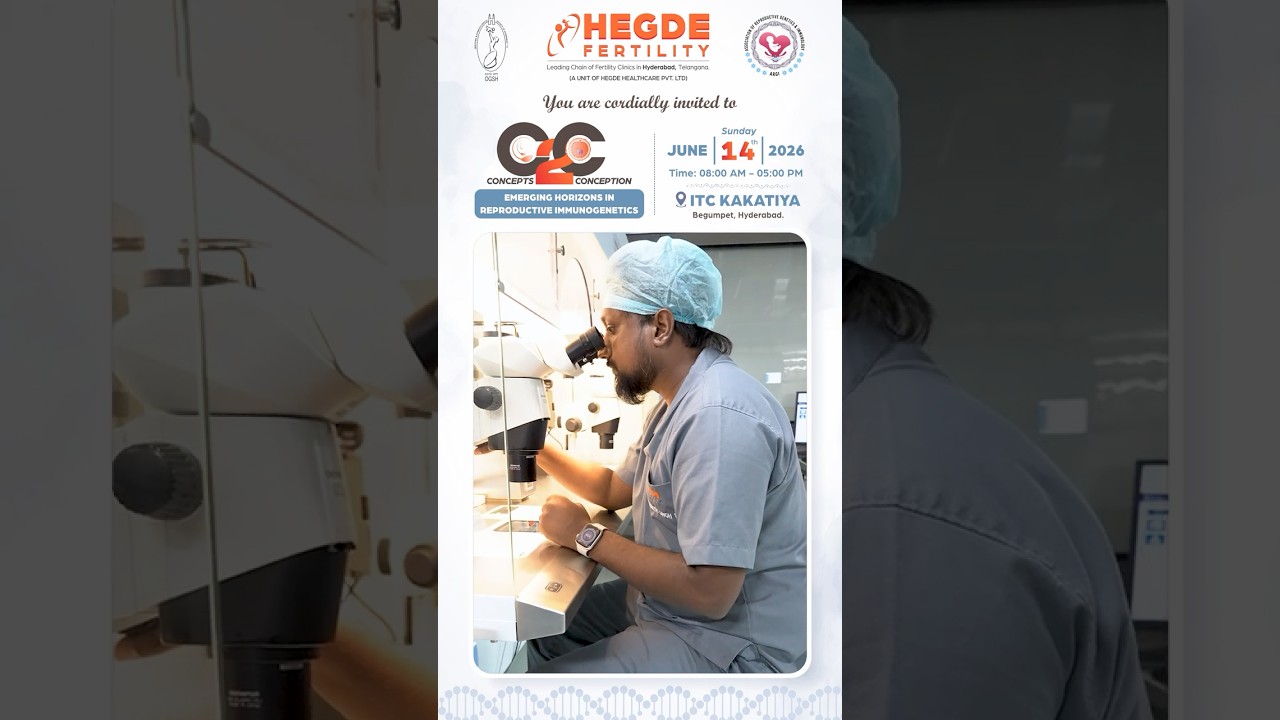
C2C | Concepts 2 Conception #Conference 2026 | Fertility Conference #C2C #Event #ReproductiveHealth
Hegdefertility
891 views•2026-05-28

Koji - the enzyme powerhouse 💪
EdibleAlchemy
18K views•2026-05-31

KPV Peptide Benefits
ReganArchibald
168 views•2026-05-29
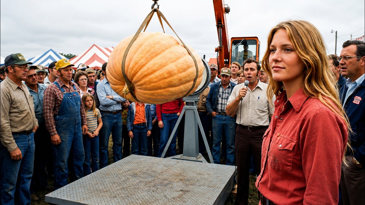
A Paper Mill Dumped Wood Fiber on Her Farm for Years...She Used It to Grow 800-Pound Pumpkins
FarmlandChronicles
436 views•2026-06-02

The Prague Chimera – What We Know So Far and Our Experiments
themulberries
619 views•2026-05-28

Every Genetic Gift You May Have Explained
ChefCalebYT
211 views•2026-05-31

Mechanical Characterization and Modelling of Tissues (Intro Video)
npteliitd
109 views•2026-06-02


