Dr. Swain delivers a rigorous and well-structured synthesis of neurophysiology that effectively bridges historical discovery with modern anatomical detail. It is an indispensable resource for students seeking a clear, foundational mastery of synaptic communication.
Install our extension to search inside any video instantly.
Synapse: The meeting point of two Neurons: Understand in 20 minutes
Added:Synapse a structure which is said to be the meeting point of two neurons. It is the structure which connects the neurons of the body and it is the structure which decides how electric signals will communicate from one neuron to the other.
Today this lecture is about the syninnapse, its structure, its history, how a typical synapse is formed and then we will learn how the synaptic transmission occurs. Therefore, I have divided this lecture into two parts.
Today is a basic introduction as well as history to the synapse. Literally speaking, the term synapse is not new, but its organization and structural forms are completely new with the advancement of the science. In 18th century, the Golgi was the person to develop a stain and he used the silver staining to stain the neurons. After that, Kazul was the man who used Golgi's staining method and said that neurons are structural and functional unit and they are separate from each other. But it was 1897 when Charles Scott serrington was the person to coin this word synapse literally meaning the meeting point of neurons or handseacking between the neurons. But surprising to this 1921 is the year in which the synap and synaptic transmission was discovered. It was the fight and debate between the neuroscientist that whether the transmission across the synapse is electrical phenomenon or a chemical phenomenon or a combination of electrical or chemical. But it is a famous experiment of Otto Louie who is said to be the master man behind the concept of chemical transmission across synapse. So with this very brief introduction I will tell a little about this autolo famous experiment. Students you must know how the history of the synapse has been developed. So it was Golgi then Kajill and after Kazil Serington was the person to coin the word synapse and thereafter the famous classical frog heart experiment of autoloi and therefore that word synapse what we are today understanding the concept of synapse in the form of chemical transmission that is the pioneer work of autoloi and later on it was supported by Dell according to them Dell and Olloy that neurotransmitter discover discovered was acetylcholine.
In 1950s there is a discovery of electron microscopic structure of synapse and then this synaptic organation came into picture. Later parts of 19th century and 10 20th century it is a remarkable period to understand how these neuronal synapse they function and what do you mean by plasticity? How neurons get developed, modified with respect to the external stimuli and modulate themsel for functionality that comes in later parts of 20th century. And now with the advancement of science, imaging techniques, electrphysiology and implanting technologies, it is very much apparent that how neurons are functioning and our body system along with the synapse function. Without the synapse, body cannot work. The process of learning, the process of modulation of brain behavior, brain communication system, motor activities, everything what you are seeing it is a it is due to the synapse and synaptic function. So let us understand how this synapse is organized and what are the fundamental parameters in this introductory lecture.
So before going into the details, please do not forget to like and subscribe my channel.
So let us beginning with this word synapse which literally meaning it's a physiological structure structure and it's the meeting point of it is the meeting point of two neurons.
It's a classical term was coined by this great man Charles Scott Sarington and he is known as the person behind the concept of the syninnapse.
Second, the syninnapse source chemical transmission chemical transmission and this was confirmed by the very famous experiments of the scientist Ottoi that is in 1921.
This is 1897.
Loi performed the famous isolated frog heart experiments.
These are classical experiments of neurobiology.
It was well known that the heart rate is reduced by vagus nerve stimulation because vagus nerve releases certain chemical which causes reduction in the heartbeat. I'm telling about 1921 story.
It was not known whether that chemical is known as acetylcholine but it is well known that electrical stimulation of the nerve reduces the heart rate. So what Ottolo performed you cannot believe students that Ottolo saw a dream in the night and in the dream he designed this work and then he laid down the concept of setup of experiment and then he carried out the experiment and it changed the entire understanding of the synapse and synaptic biology. So what he performed now he took two heart from frog and he kept in two chambers or beers you can say in one of the heart the Vegas nerve is intact and in other the nor was caught. The Vegas nerve was caught.
Both were kept in salt solutions which will support the heart function.
I'm explaining this experiment in a very simple language to draw your attention how this concept of chemical transmission was discovered. So what they what Louie performed he simply stimulated electrically.
He stimulated electrically this vagus now and he marked that the heart rate is decreased which is well established. Then what he did he simply took out some of the fluid from here and pour into the heart containing the chamber without the vagus nur and what accidentally he marred. His observation was that the heart rate was decreased.
Then Louie considered that here the vagus is intact but here the vagus is already caught. Then how putting this chemical will reduce the heart rate.
Then Otolo came out with a very beautiful concept that when he stimulated this as some chemical he was not knowing the name that chemical reduced the heart rate and that chemical is there in these solutions. From this perfusion chamber when he moved that solution to the other perfusion chamber he marked that there is lowering of heart rate. That means that chemical is present here in this fluid or in this fluid which when poor it caused the heart rate to decrease. So this was the first classical experiment in neurobiology which stated that synapse shows chemical transmission. And Dale was a person that Hel in 1939 he coined that word acetylcoline means he he just simply said that was right saying that this chemical transmission occurs. So thereafter this word chemical transmission in synapse came out. That means a synapse is a structure anatomically it is not connected. It maintains a space of around 20 to 200 to 300 anstrong. You can say that space is known as synatic cliff. And here there is no physiological connection is there.
Then a simple question comes to the mind. If if you see a structure like this this is one neuron. It is showing the end points. I will tell you the detailed structure and it is another neuron and this one is the space that is known as the synaptic cl. Then question arises that if the impulse is coming from this side, how this impulse will come to this side means how neural transmission will occur if there is a gap is there. So this clearly justifies that there are some anatomical and functional functional modalities are there which are favoring the process of transmission of impulse. So let us understand this structures in detail then only you can understand how this occurs. Now coming to the second part how this conduction is said to be unidirectional.
That means if the impulse is coming from this neuron it will go to this neuron but it will not go back. So why this occur? So that is a very interesting question why synaptic transmissions are unidirectional. Now third question which comes to my mind whether all these synapses are chemical synapse or whether there is any other kind of synapse are there basing on this electrical event.
Answer is two types of synapse are there on the basis of modality of conduction.
The number one is the commonest type.
More than 99% of the synapses of our body are characterized under this category which are known as chemical synapse where one impulsive will come from this side. It will cause the release of the neurotransmitters in this area. They will bind with this post neuron and there will be conduction.
This type of synapses are chemical synapse. Students we'll deal in detail in my coming lecture. Number two point, there are some neurons are there or some synapses are there where they are in contact with each other. That means there is no functional synaptic clap.
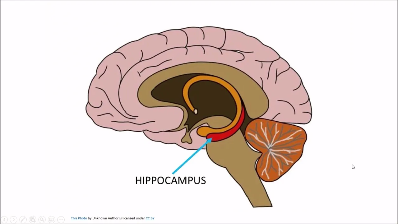
You are saying this junction is by means of gap junction and there is a direct communication of one neuron to the other. Therefore, therefore what happens the conduction becomes very fast. But for your knowledge remember chemical synapses are showing unidirectional transmission as I told but when they are connected with each other those are electrical synapses which shows birectional conduction that means conduction can go up and down and there is no delays there no physiological events occur in the synapse during the time of conduction in electrical synapse that's why conduction is very fast whereas in a chemical synapse number of physiological changes occur which we will deal in detail And that process leads to synaptic delay. That's why there is a conduction process is there which is relatively or comparatively smaller compared to that of the electrical synapse. And electrical synapses are very for they are modified for very fast processing. They are not present everywhere. Probably they are mo they possibly they are confined to the hypoc campus and some of the faster parts of the brain. Otherwise you can see the most of the synapses are chemical. The second very important point what I told plasticity it is the capacity of the synapses to modify themselves. It is the capacity to reorganize themselves. It is the capacity to reorganize functionally and structurally that is always seen in the chemical synapse but not in the electrical synapse. So with these very brief introductory points let's come to the conclusion of this particular word.
The syninnapse is a meeting point of two neurons. Physiologically they are connected with respect to the transmission mechanisms but anatomically they are not connected rather they are separated. So now I will show some other fundamental points and we'll come to the conclusion of this class and in the next lecture we'll see how a synapse is organized and what kind of morphological and anatomical and physiological modifications are there which makes it very special.
The diagram what I'm drawing this is a cryptic diagram to show the basic organization of the synapse and how the synaptic transmission occurs. So do not forget that we are going to deal in detail and this is a series of around 10 lectures where we will try to understand how neural system functions in mammals.
Okay. So what is the point? a synapse a typical synapse. So this is the literal diagram I'm showing this is axon endings as I already explained the neuron structure so you people can understand these are the bulb like structure or bon like structure or axon terminals or synatic buttons. So this is the meeting point of the two neurons where you are seeing a functional space. So what I have drawn I have drawn this much apart for your better understanding and this is the diagram which shows the typical organization of a chemical synapse. So what the diagram is showing it's showing a bulbular or a globular structure and this globular structure is known as the synatic bottle and if the impulse is coming from this side we will say it as this is the preinaptic side and this is pre synaptic exon terminox or button terminus or we can say as synaptic ball presinatic neuron because the impulse is coming from this part and it will go to next part. What it shows? It shows a definite membrane. This membrane is known as presinaptic membrane.
I'm drawing very easily so that you people can understand this is presinatic membrane.
In the presinatic ball you are seeing a large number of energy producing structure and as you know these are known as the powerhouse of the cell and they are the mitochondria which are the regulatory factor for ATP production and they are the regulators of transmission as well we will see. So these are large number of mitochondria which are present in this. So we should not forget how this is being working. So this is mitochondria. Now I'm drawing large number of vasicular structure membrane bound vasicular structure which are present in the presinaptic bulb and that makes it very very specialized and these vesicular structures are containing the neurochemicals which are stored in them so that during the time of transmission they are going to be released and these structures what you are seeing they are known as the sinic vesicle very very important structure.
These are synaptic vesicles.
Their number varies from the synapse to synapse but the more active synapse contain 100 to 1,000 number of vesicles but their number varies. These vesicles are derived from golgi complex and are processed by the endopplasmic reticulum and they are known as transjectional vesicles. They are present freely in the cytoplasm but they are not attached to the membrane and these synaptic vesicles are membrane bound that means very important and they do bind with specialized proteins and filaments which are present in the presinaptic ball that we are going to discuss and these specialized proteins make these synaptic vesicles help in the process of fusion and release of neurotransmitters. When these vesicles join to the presinaptic membrane they are known as bound vesicles and these bound vesicles they so the process that is known as exocytosis and these exocytosis lead to the release of neurotransmitters the chemicals. So here some of the vesicles come and they will attach to the membrane and thereby these vesicle fusion will lead to the release of the neurotransmitters and we will see that process in details in my coming lecture.
That process is known as exocytosis and it causes the release of the neurochemicals or the neurotransmitters into the synaptic cl. The third and the last very important point that this membrane contains the specific ion channels which are responsible for the influx of the calcium and calcium is the regulatory factor for this neural conduction mechanisms and neural transmission mechanisms. So these are nothing but the voltage gated calcium channels. So for this class I can say only this much that whenever one impulse comes these voltage gated calcium channels depending on the voltage they open and this causes the influx of the calcium from the extra synaptic space.
So the calcium will enter and thereafter the calcium will interact with the mitochondria as well as the transporter proteins which are present in the synatic bul along with the filaments so that these vesicles will move towards the membrane. There are three to four types of movement are being visible depending on the type of vesicles. These vesicles are coming from the axon surface to the somatic surface through the axon by exoplasmic flow and once they will come they are free. Sometimes they start moving when this calcium will come and they are known as transitional vesicles and these transitional vesicles once they come and fuse with the membranes they are known as bound vesicles. Guys, please remember that these vesicles are the identifying feature or the typical feature of the presinaptic bulb and point to remember these are not present in the post synaptic side and that's why I said that the chemical transmission across this is interactional because the directional transmission require the receptors which are present on the post side but they are not on the pre-side and the pre-side is predominantly characterized by these vess vesicles and besides these vesicles we are also seeing number of proteins are there contractile protein transporter proteins binding proteins and these proteins are I can say these are synaptic proteins we'll discuss in the coming lecture these synaptic proteins they regulate the transportation mechanism and they are the regulators of the synaptic vesicle flow as well as the connection to the membrane and these synaptic proteins they also regulate the presinaptive filaments those which are present in the presinaptic ball. So they therefore they are known as the regulatory proteins.
Now in the last part we can see that is the post synaptic ball a globular structure which is present here and it is showing receptors on its surface and these receptors are mostly liant bound receptors and they respond to these neurotransmitters they will come and bind here so that number of physiological electrical changes will occur that's my coming lecture where we are going to discuss but for the And for the time being we should understand that these neurotransmitters once they bind with these receptors then there is electrical activity occurs. If the electrical activity occur depolarization occur then it is known as excitatory synapse and the impulse will travel and if the neurotransmitter is binding and there is no depolarization then it is known as inhibitory synapse where the conduction is stopped there. Basic difference between the two is that once the neurotransmitter binds if it is activating or allowing the entry of the sodium then depolarization will occur and this type of synapse is known as excitatory synapse and is it generates excitatory post synaptic potential and if it opens chloride channels chloride channels are inhibitory and then the synapse is known as inhibitory synapse.
So the point to ponder is that here you are getting vesicles no vesicles here you are getting the neurotransmitters which are released to this fundamental space this is that space this fundamental space and I am saying them as the synaptic cliff and this synaptic cliff I can say 20 to 40 and strong this is the lane and this synaptic cliff is the site of release of neurotransmitters but the most and the important character is these receptors. That means we can remember that receptors are present on the post synaptic membrane and in the presinaptic bulb you're seeing the presence of the vesicles and the neurotransmitters.
So why synaptic conduction is unidirectional. Now it is very clear because of vesicles neurotransmitters in the preside and no vesicles on the other side rather receptors and these receptors may bind and stimulate the conduction of impulse. then it is excitatory. If they inhibit then they the if there is no excitation is there then it is inhibitory and chloride channels they inhibit and sodium channels they activate but we have to remember here you are not getting that much of calcium channels. Sometimes ion channels are not attached to these receptors rather some secondary messengers are attached and these secondary messengers instead of allowing the entry of the ions they will activate the groin the communist mechanism GPCR G- protein coupled receptor mechanism.
So my coming lecture is on that what these receptors bring out how these neurotransmitters are released how neurotransmitters are coming from the vesicles. So with this this particular lecture came to a conclusion that is introduction to synapse. Draw this diagram for your reference because these are the post synaptic membrane and I I can write here this is post synaptic membrane and these are the receptors and these receptors they will either allow the conduction or inhibit.
Okay. So coming to the conclusion but there are two three questions remain and just before finishing I'm putting these questions so that that will be our next lecture. Number one how these synaptic vesicles come and bind. Number two how these synaptic vesicles release the neurotransmitter. Number three where these neurotransmitters go number four where these neurotransmitters bind.
Number five how these neurotransmitters bind and what they bring out. Number six what do you mean by post synaptic conduction? Number seven, what is post synaptic inhibition? Number seven, number eight, what happens to the neurotransmitters after the conduction is over? Number nine, what do you mean by synaptic delay, synaptic fatigue? And what do you mean by summation effect, special summation and temporal summation? So all together we are going to discuss in the next class. So before leaving this class, one of my question what is the difference between the electrical and the chemical synapse?
already I have mentioned but keep these points in your written form and I will explain in the next lecture so that you can compare your answers. So thank you so much for your patient hearing. Keep learning and keep seeing my videos.
Please like and subscribe the channel.
Thank you so much for your patient hearing.
Related Videos
8:08
Neuroscience
What is neurodegeneration?
TheSheekeyScienceShow
3K views•2019-08-19
57:35
Neuroscience
IPL - Ruth Empson "Mind the Gap"
otagouniversity
251 views•2019-07-08

14:01
Neuroscience
How our body shapes our mind | Pancho Tolchinsky | TEDxNapoli
TEDx
2K views•2019-12-05
1:00:02
Neuroscience
Impact of Early Life Deprivation Danielle Stolzenberg Marcus Pembrey Bruce McEwen
uctv
4K views•2019-12-01
10:32
Neuroscience
β-Caryophyllene for Parkinson’s: Protecting Dopamine & Easing Symptoms
parkinsonsdiseaseeducation
6K views•2025-08-09

20:44
Neuroscience
New Insights from Inside the Brain with Rodrigo Braga, PhD
NUFeinbergMed
421 views•2025-04-14
9:57
Neuroscience
Highlights for dystonia • 2025 MDS Congress
movedisorder
145 views•2025-10-27
58:17
Neuroscience
Animal Welfare Synergy Series: Dr Tom Smulders on hippocampal neurogenesis
costactionlift
135 views•2025-07-14
Trending

13:25
Business
Playstation NO DISC/NO BUY Fight Is Over...
DavidJaffeGames
4K views•2026-07-23

16:52
Anthropology
Americans Confused in Australia for 17 Minutes Straight
IWrocker
17K views•2026-07-23

7:38
Blockchain & Crypto
Bitcoin Social Interest: Dozens of us Left
benjaminjcowen
12K views•2026-07-23

11:32
Business
Tesla Profits Plunge & SpaceX Stock Continues Fall
TheJohnJohnstonLounge
6K views•2026-07-23