Bacterial cells possess a three-layered cell envelope consisting of the plasma membrane, cell wall (made of peptidoglycan), and glycocalyx (a polysaccharide coating that can be flexible as a slime layer or rigid as a capsule). The cell wall provides shape and protection against bursting and collapsing. Bacterial surface structures include pili (for conjugation), fimbriae (for attachment), and flagella (for locomotion, composed of flagellin protein without the 9+2 microtubule arrangement found in eukaryotic cells). Mesosomes are infoldings of the plasma membrane that perform functions similar to eukaryotic organelles including cell wall formation, cellular respiration, DNA replication, and DNA distribution. Photosynthetic bacteria contain chromatophores (pigmented membrane structures) and gas vacuoles (air bubbles for buoyancy), while non-photosynthetic bacteria may have inclusion bodies for storage. Bacterial ribosomes are 70S ribosomes (composed of 50S and 30S subunits), which are the smallest cell organelles and can form polyribosomes on mRNA for efficient protein synthesis.
LT - 2026 - 2027 _ ONLINE BATCH _ BOTANY [ SUMAN sir ] CLASS -08
Added:Okay, coming.
Voice is coming. Check it out.
Hello.
Is my voice audible? Come on.
Come on, students.
Okay, done. See around the cell there should be a plasma membrane. Then second one is cell wall.
Around the cell wall some bacteria possess some extraordinary structure or some kind of extension. That extension is known as glycoalics.
In this three layers of envelope has been developed around bacterial cell.
These three layers of envelope one by one if you check first one plasma membrane second one cell wall and the third one is known as glyco callex the three layers these three layers collectively known as cell envelope Three layers are collectively known as cell envelope. Let us talk about them one by one. First one, what is the story of glycoallex?
Glyco means carbohydrate. Callex means outer covering or rigid covering. That's called glycoallex.
Glycoal is a kind of polysaccharide coating.
Polysaccharide coat around some bacteria. Not all bacteria possess glycoalics.
Most of the bacteria without glycoalics they can survive. If a bacteria possess glycoalics, it is made up of polysaccharide. Next one. Glycoalics will be on the basis of its physical composition. Sometime it is flexible.
Flexible.
Sometime it is tough and rigid.
Very rigid and tough. If it if it is a flexible layer, the glycoalix is having a name called slime layer.
Slime layer.
Next, if it is a stuff and rigid that's called capsule.
Listen, these things we have come across in molecular basis of inheritance.
If you recall it very closely when we were talking about molecular biology transformation principle that was discovered by Griffith we had discussed there Estrain A strain type of steal pneumonia we have mentioned there Estrain is virulent arin is nonvirolent what is this estrain and arain those which are having the glycoalics are called S strain. Those which are not having glycoalics are called R strain.
Simply to remember in coordination to that information generally those bacteria which are having glycoalics are virulent bacteria. Many bacteria which are having the glycoalics are capsule or slime layer. They are virulent in nature. Glycoallex bacteria virulent are pathogenic in nature pathogenic this one is there in our NCR book that's why I'm saying but it doesn't mean that every bacterium which is having glycoalics definitely would behave like a pathogenic but generally pathogenic bacteria are having glycoal coat on the top of them Okay, then come back to next one. Cell wall.
Cell wall.
[snorts] First question. Do all bacteria will have cell wall?
No. There is a group of bacteria known as myopplasma.
Myopplasma is a bacteria or it is the bacteria without cell wall. They don't have cell wall. Which one? Myopplasma. Very good. If they don't have cell wall, how could they survive? Be careful. In order to survive, it is not compulsory to have cell wall.
Cell wall is there. It gives some advantages.
It doesn't mean that a bacteria will survive only by the presence of cell wall. That's not the thing. Then come back. Cell wall if present made up of what? Made up of pepidoglyon.
Pepidoglycon commonly known as murine murine or pepidoglycon.
Next one it is a rigid layer. First thing shape of cells shape of cell is due to cell wall shape.
We have seen bacteria possess variety of shapes like basilus rod shape. Coke, spherical, vibrio, comma, shape, variety of shapes will be there. All those shapes are developed due to presence of cell wall. Cell wall is the reason behind shape of the cells. If so, earlier you said something called myopplasma and you have denoted myopplasma do not have cell wall. Does it mean that the myopplasma will not have any shape? Of course.
Okay. My plasma pleomorphic in nature pleomorphic in nature. Pleomorphic in the sense they don't have proper shape. The bacteria that does not or a cell that does not possess any proper or particular shape.
That bacteria is known as pleomorphic bacteria. Next one. This also I mean cell wall protects the cell from bursting bursting and collapsing bursting and collapsing. What does it mean by this bursting and collapsing?
Imagine I have taken a cell kept in hypertonic solution. That means around the cell the concentrated solution is present. Whatever the water present in the cell started to leak out. As a result cell shrink that shrinking process is known as collapsing. In addition to that, if you keep a cell under hypotonic solution, that means water-rich solution, water started to penetrate inside the cell. As a result, there will be a a kind of swelling will happen inside the cell. That's called bursting or eventually cell is going to burst. Like this cell neither burst nor collapse immediately due to change in external environmental solutions. That would be the protection given by cell wall. Then go to most important plan part that's called plasma membrane.
Now one thing about that whenever we see plasma membrane plasma membrane is the universal structure for a cell. In fact without plasma membrane there is no cell. So do all cells have similar type of plasma membrane?
almost same with some minor additions and subtractions will be there in the nature of chemicals present in the plasma membrane. But almost all kinds of cells will have same structural orientation in the plasma membrane. that indicate structurally similar to structurally similar to ukariotic cell plasma membrane. That's why whatever the details that we will study about plasma membrane, we will study about one time about plasma membrane. Okay. And here after cell envelope primary discussion we should talk about one matter that's called gram staining.
Gram staining there was a scientist called Christian ghed chemical or a chemical stain on the top of bacteria called crystal violet.
Crystal violet commonly known as g stain.
He had taken few drops of crystal violet added on the top of bacteria.
Some bacteria had absorbed this color.
Some bacteria couldn't be absorbed the color. The bacteria which absorbed color so-called crystal violet color he named them as gram positive bacteria. Some bacteria couldn't absorb the color and named it as gram negative. Simply if crystal violet absorb g positive they intake or absorb the color.
They can intake or absorb color. Gram negative bacteria.
Gram negative bacteria cannot absorb color.
Cannot absorb color. That altogether makes gram positive bacteria will have thick cell wall. The cell wall is very thick. Gram negative bacteria is having the cell wall that is thin.
cell wall very thick in gram positive very thin in gram negative bacteria.
This might be clearly understood like this. When we add few drops of crystal violet those bacterias which are having thick cell wall can absorb that color retain the color for future purpose or else in future the color will be present in the cell wall. But some cases immediately after absorbing the color a cell wall could not retain that color for future. This we will call it as gram negative. The earlier one is called gram positive. Altogether thick cell wall containing bacteria are can retain the color called gram positive. Thin cell wall containing one cannot absorb the color. This is ground staining procedure. I did not explain the procedure of ground staining in completely but this much information fine to understand the information that they have given in our NCR that much fine. Then go back to cell. On the surface, what are the structures? We will see on the surface of cell envelope we will see structures like flaga.
Flazilla.
Next one we will see some structures like pi.
Pi.
These are pi.
fili and some more structures like fimria.
The way I am representing these structures on the top of the bacteria, it is essential. Three extensions will be there on the top of the envelope. One is flazilla, pili, fimri. What are those extensions and their usage? First one I will talk about pilli.
If you take pilli which is also known as sex pili.
Why? This is a kind of tube or tubular structure help in conjugation process.
Simply to understand in bacteria there are some modes of sexual reproduction. Exact mode of sexual reproduction may not be identified in bacterial cells like meiosis, fertilization, gyote formation.
These events are not there. But procariotic cells do have their own mode of sexual reproduction in order to develop variations.
The sexual mode of reproductions are conjugation, transduction, transformation types. In that conjugation is a kind of physical association of two bacteria.
Two bacteria will come close to one another and they will join by formation of a conjugation tube. That conjugation tube is formed by pilli since it is helping for sexual reproduction in bacteria. That's why sometimes it is called as sex pilli. Very important.
Apart from that pill has no other role to be conduct. Next one more is there fimria.
Fimria are bristle-like structure. Bristles.
Bristles means small hairlike structures.
What is the bristle-like fimria work?
They help in attachment of bacteria on solid surface or on substratum.
Generally this fimria will be present on those parasitic bacteria in order to make them to attach. I mean the bacteria has to attach on the surface of host cell to make itself cling or attach to the host cell. The bacterial cells develop some special kind of briilike structure that is fine hairike structures. These hair like structures are known as fimria.
Apart from that fimria also do not have any work. Next one. PI made up of a protein called fillain. Both are protein structures.
Protein. Fillain.
Fimria made up of fimbrin.
Either you take fili or fimri they are made up of proteins but one interesting common thing about them both [clears throat] do not work in locotion not useful for locom motion.
Locom motion of bacteria is not possible due to pi or fimria.
Now how does a bacteria can be motile?
Careful in a ukariotic cell there may be so many varieties of locom motor organs are there. In fact ukariots will have flazilla celia and in rare cases pseudo podia may also help in locom motion but a procariotic cell will have only one kind of locomotive organ that's called flagula.
Flazilla.
Flazilla plural. Plazylum singular.
Fazilla present in locary or those bacteria which are able to be motile all will have one thing that's called flazilla nothing else. Imagine there will be some bacteria who cannot motile. There will be some bacteria who are motile.
All motile bacteria possess flazylum.
Plazylum is the one which is exclusively belongs to motile bacteria. You have seen a bacteria with flazilla.
That bacteria definitely can able to show motility on its own. Next one. The flazilla made up of made up of flagillin protein. It is [clears throat] also made up of flaziline protein which means 9 +2 microtubule arrangement absent.
9 +2 array of microtubules is absent.
Why do you say like that? Simple. When we talk about ukarotic cell, a ukarotic cell, flazilla made up of microtubules.
Microtubules are exclusive characters of ukariotic cell. In that case in case of procariotic flula motility and characterization may be same but internal structural orientation is completely different. Procariotic cells will have flazilla simply made up of flazilin protein. But in ukariotic flazilla microtubules are present which are arranged in the form of 9 +2 arrangement or 9 +2 array of microtubules will be present. Keep it in mind. Later when we will talk about end of the story in the cell the unit of life we will have flazilla structure in ukarots and also centrosome structure those structures we will in detail discuss but altogether whenever we talk about procariotic flazilla. Proariotic flazilla is made up of flaziline protein but that protein never follows any one particular mode of arrangement of microtubule like structures. The just protein filled large filament will be present. In this case procariotic flazilla will have three structures.
Flazilla posess three structures. One is [snorts] basal body that is the where flazilla originated.
Next one hook. Very good. Next one followed by filament.
filament in this case. [snorts] First one if you talk the basal body where does it present the basal body present in plasma membrane within the plasma membrane there will be a presence of basal body this is the basal body for example actually from plasma membrane basal body originated gradually it will cross our cell wall and also So if glyicoal present glycoalics also but overall the basal body will comes out of cell wall that's why always this flulilla is started to appear it is coming from cell wall but actually flasilla will come from plasma membrane.
Origin is plasma membrane. But externally if you see the plasilla started to appear like it is comes out of cell wall. Because if you see from the outer surface first you will see cell wall then it appears like something is protruding away from cell wall. That protruding body we can call it as flazilla. But actually plasilla would be originated from plasma membrane because the basal body will be present at plasma membrane only. Cell wall something dead which could not be generate anything.
Cell wall itself is secreted by our plasma membrane. So if something is growing on the top of the cell that material must be secreted by our plasma membrane. So on the cell wall there is a presence of basal body that basal body will also having hook then followed by filament. This is the filament.
This one is hook and this one is basil body.
These are the parts. Next thing do all bacteria have flazilla? No. What type of bacteria will have flazilla? Simply whatever the bacteria which can able to motile that bacteria should be having flazilla. Flazilla has no other work at all. They have only one work that's called loco motion. Keep it in mind. But another thing location and number of lazula number of flazilla varies from cell to cell.
location as well as number of flazilla varies from cell to cell. Some bacteria might be having single flazilla at one end. Some bacteria may have two flazilla at the opposite end. Some bacteria might be having a bunch of flazilla at one end. Some bacteria will have flazilla all over their body. Like that number distribution may vary from cell to cell but on in all there will be a variations in the number of plasilla and also the location of plazilla present inside a bacterial cell that's about the flular arrangement. Then after the flazular structure let us see what they are talking other structures in internally areas. You will see the plasma membrane of bacteria will started to infold.
Plasma membrane started to infold. These infoldings of the plasma membrane are known as misosomes.
Misosomes. Let us learn some details about misosomes and their importance.
First one, who are misosomes?
Misosomes are unique to procariotic cell. I'm not saying all procariots will have misosomes. That's absolutely wrong.
Some of the procarotic cells do not have misosomes. But if misosomes present what is their orientation and their functions? See misosomes are infoldings of plasma membrane.
Misosomes are infoldings of plasma membrane. What do you mean by infoldings?
Plasma membrane itself will folded towards inner side and it is making something is called misosomal structure.
In that case shape will be tubular.
Sometimes tubular tube like structures like this tube like structure sometimes they are vesicles vicular structure like this. Sometimes even they are lame.
Lamlay means membranous sacks called lamele like that shape of misosomes may vary from cell to cell they don't have a constant shape because they are infoldings of plasma membrane then important thing to be noticed their their functions functions of plasma misosomes first and foremost function cell wall formation If you recall cell wall of the bacteria made up of pepidoglycon or murine that meine or pepidoglyon chemical synthesized by plasma membrane and it would be secreted outside that's why cell wall formation is under the control of our misosomes.
Next one. Now compare this one. If you could recall what was the cell organal in a ukariotic cell especially plant cell responsible for cell wall formation that was goli complex. Golgi complex in ukarotic cell will help in cell wall formation and in [snorts] case of procariotic cell misosomes responsible for the same cell wall composition. Next one.
They also help in cellular respiration.
Cellular respiration.
Don't mistake respiration means exchange of gases.
Now exchange of gases means breathing.
What about respiration?
Respiration is a technique inside the cell where food get oxidized in order to release energy in the form of ATP that is called as cellular respiration. Okay.
Respiration require gases. That's why multisellular organisms can have some separate variety of respiratory organs only for the sake of breathing. Come back to the story. If you are talking about misosomes at the site of cellular respiration, what does it mean by that?
They are responsible for synthesis of ATP.
Synthesis of ATP. When it comes to the word ATP, you can directly correlate in a ukariotic cell the ATP missionary is mitochondria. Obviously more mitochondrial function more ATP can be synthesized by a ukariotic cell. Now the question under our level does the procariotic cell required ATP?
Of course no living cell can survive without the requirement of ATP.
If mitochondria is absent in procarotic cell, there should be a someone to be compensating that loss. That someone is nothing but misosomes. If you closely observe mitochondrial function, cellular respiratory events like link reaction, KB cycle and also electron transport chain.
Link reaction, KB cycle in the matrix.
KB link reaction, KB cycle in the matrix. Electron transport chain might be happening on inner membrane. That inner membrane electron micros electron transport chain. Do you think is it possible in procarotic cell because they don't have mitochondria?
Of course possible. that would be happen on membrane of misosomes. Be very careful.
Almost all activities which are conducted by a ukariotic cell, a procaryotic cell can also do. But the different places in the different locations it is possible in case of procariotic cell. If anybody ask you a question does a procarotic cell can conduct electron transport chain as like the mitochondrial inner membrane. Of course mitochondria is absent but misosomes are compensating the work of mitochondria in a procarotic cell. Very good. Next one. Another work of misosome that is DNA replication.
DNA replication.
Now I wanted to say one thing about DNA replication.
Recall how much time a typical bacteria takes for the cell division or binary fusion.
Simply it will take around 20 minutes only. In that 20 minutes bacteria spends or bacteria spend 18 minutes only for the DNA replication.
If you could remember I said bacteria process circular doublestranded DNA that circular doublestranded DNA is present in the form of nucleoid or naked DNA.
That naked DNA is not always freely present in the cytoplasm.
Mostly it is associated with misosones.
Why it has to be associated?
The quick way of DNA replication is possible during at the time of cell division or binary fusion with the help of misosomes.
Whatever the enzymes required for the DNA replication, all the enzymes would be provided by our misosomes. As a result, bacterial DNA can be replicated.
Misosome function not only ends to support DNA replication.
Even after the DNA replication, the misosomes will support the replicated DNA equally distributed to two daughter cells. What does it mean by that? Let us see one thing. Distribution of distribution of replicated DNA replicated DNA to daughter cells.
Why are you saying this? This is very important. Many people I have seen constantly misunderstanding what does it mean by distribution of DNA equally to two daughter cells. Let us see in a ukariotic cell how does distribution occur.
If you consider a ukarotic cell in a metaphase there is a clearcut development of two chromatids in one chromosome. These two chromatids each chromatid contain one DNA that DNA has to be entered in future daughter cells.
There will be a phenomena called spindle fiber attachment on each centromeir both sides you know these things that's why I'm simply moving there is a centromeir both sides of the centromeir one side here one kinetto core another side another kinetto core will be present for both kinyottoes opposite poles spindle fibers will come and they will attach during the na phase is they will drag these two chromatids away from each other. As a result, centromeir split chromatids separate and each chromatid will move to opposite pole. As a result, the distribution of DNA that was replicated during Sphase possible in case of ukariotic cell. This much long story is not possible in procarotic cells. Why? There is no concept of spindle fiber formation. If you recall the procariote cells undergo amitosis which means there will be no formation of motic threads. Motic threads are absent in case of procarotic cell. So there should be a some way we can give that replicated DNA molecules equally to two daughter cells. That way is through misosomes. Misosomes are the one who will split the replicated DNA and given to two daughter cells equally.
This is the reason why misosomes help in distribution of replicated DNA equally to two daughter cells. Am I clear the word? Next one. One more function cell secretion.
What does it mean by cell secretion?
Anything that should be goes out of the cell in case of ukariotic cell the secretion is under the control of a special cell organal called golgi complex. Later we will talk about it with respect to goli complex. Wherever a cell secretes something away from the cell anything that goes out of the cell that is strictly regulated by goli complex. But in procarotic cell gi complex is not there. So somebody should be there to compensate that work. That work is done by our misosomes also.
Okay. I mean misosomes do that work also. So observe the statement here.
Cell wall formation, cellular respiration, DNA replication, replication DNA distributed to daughter cells and also cell secretion. All these activities are under the control of misosomes.
Misosomes if not present there may be different types of functions conducted by different places but misosomes if present they are the one who do all these activities. Next one. Apart from the misosomes, some other things we need to talk about a procarotic cell membrane. On the membrane here and there we will see some parts which possess pigmented area. Pigments.
Okay. Those pigmented areas in plasma membrane are known as chromato force.
They have a special name called chromato force. Who are this chromat force and what is their story? Chrommo means chromo means color.
Let me take the chromato force story for us.
They are present on membrane. Be careful. Chromato force are not freely present in cytoplasm.
Chromato force present on membrane.
They are membranous areas. In that case what kind of structures do you call as chromato force? Chromato force pigmented membranous structures.
pigmented membranous structures. They are pigmented membranous structures.
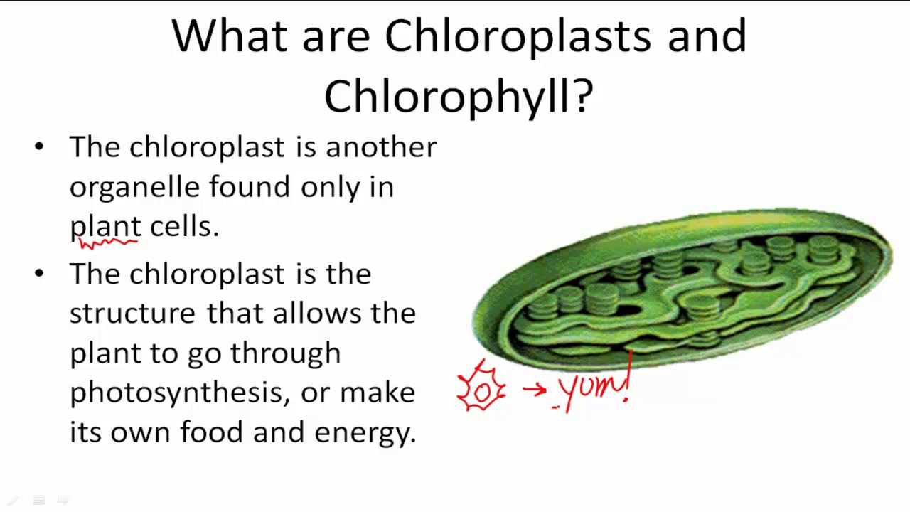
What does it mean by pigmented membranous structures on the top of the plasma membrane here and there? some amount of photosynthetic pigment will be deposited here. Very interestingly you have to remember those pigments that we will see in chloroplast will not be present in chromat force. Typically a ukarotic chloroplast consist of that chlorophyll a b and also kerotenoids jantoils but chromat force do not have all of them they have special that is chlorophyll chlorophyll chlorophyll here also rarely chlorophyll a present in case of blue green algae remaining bacteria will have bacterial chlorophyll. It's a special one. I will discuss about the bacterial chlorophyll and chlorophyll differences when we begin the photosynthetic missionary. But keep it in mind. Normally bacterial chlorophyll we will present on chromato force. In addition to that there is another kind of pigment called ficoillins or some accessory pigments. Ficoillins will be there in syinoacteria ficoins in syinoacteria cyano blue green algae we would see them blue green algae are proariotic bacteria. Okay. In that except this you don't see kerotenoids jantoils don't conrue or mistake by just listening chromat force contain photosynthetic pigments not normal photosynthetic pigments as like our plant cells possess. Plant cells will have different composition of photosynthetic pigment and bacteria or procariotic cell will have different composition of photosynthetic pigment.
That's why I'm emphasizing that. Anyhow if a ukareotic cell performs photosynthesis that is due to chloroplast.
If a procariotic cell perform photosynthesis that is due to chromat force because a procaryotic cell does not possess chloroplast still they can maintain photosynthetic missionary. Next one inside a procarotic cell cytoplasm we would see some bodies called inclusion bodies.
Inclusion bodies. Who are this inclusion? Why did you say they are inclusion bodies? Inclusion bodies means they are freely present in cytoplasm in a disappeared state. That means how many present? Where they are present? You cannot significantly notice them. That's why they are calling them as inclusion bodies. Okay. Then what is their main work? These are non-membranous non-membranous storage granules.
Non-membranous storage granules. They just meant for storage. It doesn't mean that what they will store only particularly. Anything that will be stored inside the cytoplasm of procariotic cell generally noticed as inclusion bodies.
My dears remember I have seen one statement in NCR book. You could also see something is unique present in the form of inclusion bodies in procariots.
Be careful. If they ask you procariot cells only will have inclusion bodies, that is absolutely wrong.
Why? They say procariot cells will have something unique in the form of inclusion bodies.
There are inclusion bodies in procariotic cell. Such kind of inclusion bodies we do not see in ukariotic cell.
That is the intention.
Other than that inclusion bodies are not unique to procariotic cell. You would see this statement in NCR book like this.
Something unique present in procariotic cell in the name of inclusion bodies.
Something unique that means this thing slightly different from that of ukariotic cell. If you come across a statement like inclusion body is exclusively present in procarotic cell absolutely wrong. Why?
Inclusion body is not something that is any cell organal. Anything that it is included or inclusively present in cytoplasm you can have inclusion body title. Okay. So come back to the inclusion bodies that we would see in procarotic cell. Over here there are some phosphate granules.
Phosphate granules.
Some bacteria store pro phosphate.
Some bacteria store sulfur granules.
Some bacteria store iron granules.
In addition to them there is a presence of glycogen granules. This is very important.
Glycogen granules.
Glycogen.
What does it mean by this glycogen granules?
I think a fundamental statement we know plants store food in the form of starch.
Animals store food in the form of glycogen. This we are studying from eighth class or seventh class onwards.
Now here the question will arise other than animals is there any member store glycogen?
We don't have just only animals in this world. In addition to animal we have fungi, we have bacteria, we have protozoans, protista variety are there.
So in addition to animals, is there anyone store glycogen? Okay, keep it in that question in the mind. Next one, in addition to plant, is there anyone store starch?
If you closely analyze these questions, generally other than plant remaining store glycogen, glycogen is a commonly storage food material. Glycogen stored by bacteria, glycogen stored by fungi, glycogen stored by protista member. Wherever you come across a body that it is trying to store food especially in the form of carbohydrate that food will be stored in the form of glycogen.
But when it comes to plants plants are different things. Plants store starch because starch would be stored in dry form. dry I mean water deficit form carbohydrates can be stored that water deficit form is known as starch water required for other kind of carbohydrates to store that water required components are called glycogen in our body we will always store glycogen bacteria store glycogen fungi store glycogen proteasa store glycogen but plants store starch that's why they have emphasized In our lower class books, plants store starch.
Plants store starch. Why?
Because plants only store starch.
Remaining all are storing glycogen.
Next one. Bacteria also store glycogen.
In addition to this, there is some other information we need to learn. Gas vacules.
Who are these gas vacules and what are their story? Observe.
When you talk about the term vacule, did you see the vacule term only in our bacteria? No. Earlier we have seen there is a name called vacular membrane which is called tonoplast. Inside the tonoplast there is a place called tonoplasm and contractile vacule. And also there is a vacule called you know in case of amoeba you have to remember that is contractile vacule and also food vacuum.
If amoeba perform or any other protogenone has performed fagocytosis that food stored in the form of vacule that vacule is known as food vacule and some protista members will have contractile vacules. The contractile vacules will help in osmo regulation water maintenance. Getting the idea? So altogether wherever you come across generally vacule don't include that vacule gas vacule vacules you should remember three sap vacule that's tonoplast sonoplasm concept next one contractile vacule help in osmo regulation one more vacule is known as you know food vacule over there you should never include something that is called gas vacule why Gas vacules are air bubbles.
Air bubbles covered by covered by proteinrich membrane.
They don't have membrane in fact or proteinrich layer.
That's called gas vacules. Gas vacules unique to unique to proariotic cell. You will never find gas vacules in any kind of ukariotic cell. Somewhere else if you find gas vacules be careful that organism is nothing but a proarotic cell because in a ukariotic cell you will find three types of vacules. Sap vacule, food vacule, contractile vacule, nothing else in ukariotic cell. Now procariotic cells will have gas vacuum. They are unique to proarotic cell. Not all procariotic cell only in only in photosynthetic procarotic cell.
All not all procarotic cell only in photosynthetic procarotic cell. What is the relation between the photosynthesis and also the presence of gas vacule? You are saying here photosynthetic procarotic cells are having gas vacule.
Extraordinary. Okay. We believe what is the relation between a gas vacule and a photosynthetic bacteria? Simple.
In order to constantly exposed to the sunlight, some bacteria stay on the surface of the water in aquatic environment.
In order to keep themselves on the surface of the water, they have to stay on the water. Should not be submerged.
In order to protect itself from the submerging, these bacteria have developed some special adaptation called gas vacule. They have a bubble. That bubble which will have lot of air inside. That air will make this cell to make bion are floating. Help in help in bionc help in bionc or floating like that. The gas vacules are very much essential for the bacteria in order to maintain constant exposure to the sunlight to conduct the photosynthesis. Very important. Gas vacules are exclusive for only those bacteria which can able to perform photosynthesis. Could you name some examples of photosynthetic bacteria? Well-known one. Give me very good cyanobacteria.
Cyano bacteria like this we have variety of structures inside a procariotic cell. Next in addition to them we do should not forget something in the name of ribosomes.
You know ribosomes.
What type of ribosomes could you see in a procarotic cell?
They are called 7TS. Okay. Done. I'm discussing about 70s ribosomes.
Chemically.
Chemically ribosomes made up of rna plus proteins.
Ribosomes made up of rna plus proteins.
Nothing else. They have this one. You have already seen in your molecular basis of inheritance during translation process ribosomes made up of rna and also 80 different types of proteins 80 the number also mentioned there that's for 70s only they have indicated next one because they have rn and proteins altogether they are called ribo nucleio proteins. Ribosomes are known as riboucleoproteins because rn and proteins are present in them. Next one if you talk about the ribosome so-called 70s ribosomes S stands for Swedberg unit. You might be know that the Swedberg unit is a unit for sedimentation coefficient. Now the 70s ribosomes if you take they have two subunits a larger subunit known as 50s the smaller subunit known as 30s.
Remember this larger subunit smaller subunit in a non-working or inactive state they present as separate bodies. There is a ribosome floating in the cytoplasm.
catch it. Allow it for sedimentation coefficient. If it has given 30th 30th sedimentation coefficient then this is smaller 50s some other will give. Now whenever these two are joined careful during working working in the sense during translation larger subunit and smaller subunit will join before and after translation they will be present separately.
By chance you have got a ribosomal subunit which is participating in translation. At the time if you sediment them you will get 70s. That's the reason many are confusing this is 30 that is 50 joining 80 but you have written 70 now 70 is sedimentation coefficient okay it's not about subunit combination when they join and I will use it for sedimentation process allow it for settle down then it will settle down at the 70s area if I individually allow it it will settle down at 30s area this one will be settled down at 50s area but both are combinely joined and then settle down I will get 70s area both of them combinely diameter or width of the larger and smaller subunit together it is around 15 nanome length would be around 20 nanome why are you mentioning like that 15 by 20 why The reason is very interesting. These are the cell organals which are smallest in the cell. If you find out any kind of cell organal that would be very minute and smallest. You can see that units they are nanometers not even micrometer. They are in nanometer range. That's why they wanted to show these are the smallest cell organals.
smallest cell organals inside the cell.
Out of all the cell organals whatever you will come across 70s ribosomes are the smallest cell organals in the field. Okay. Then another important thing about them whenever they work I mean ribosomes work is nothing but what that is protein synthesis.
Whenever they are doing protein synthesis they have an interesting behavior like this they will find one mRNA in procarots I'm talking about one mRNA on one mRNA several 70S ribosomes will join several 70S ribosomes will join several many 70S ribosomes would get attached to a single mRNA.
Many ribosomes on single mRNA this condition is known as polyome polyome or poly ribosomes.
Now this polyome poly ribosome generally developed by procariotic ribosomes or 70s ribosome.
In fact to say wherever you could see there is a presence of 70s ribosomes in all those places there is a chance of development of polyome or poly ribosome wherever I mean to say this 70s ribosomes you will find in the matrix of mitochondria and also in the stroma of the chloroplast and in different places wherever you identify The 70ths ribosome they do this work on one mRNA several 70S ribosomes will attach and they involved in different levels of translation events. Such kind of arrangement of many ribosomes on single mRNA condition is known as polyome or polyosome. Next one in a procarotic cell where this ribosomes are found. This is also another interesting thing very very important when you talk about okay 70s ribosomes they are attached to plasma membrane of they attached to plasma membrane of procariotic cell they attached to plasma membrane of procarotic cell. Observe very closely. Why you are specially talking about this?
When we talk about ukariotic ribosome so-called ATS ribosomes, where do you think that ATS ribosomes could be seen commonly? on the top of endopplasmic reticulum in the name of rough endopplasmic reticulum that is ATS ribosome but there will be a presence of procariotic ribosome so-called 70s would be attached directly to the plasma membrane it doesn't mean that they will be present only on the plasma membrane some freely present in freely present on cytoplasm.
What does it mean by this? Somewhere else we have seen one question in pre in previous papers.
There is a presence of ribosomes on the misosome. That's correct. Misosomes also can carry ribosomes. There is no exception for that. Okay. So 70s ribosomes either freely present in the cytoplasm or they may be present in a status where they directly go and attaches to plasma membrane. Okay. Up to this whatever the discussion I have made this one is belongs to procarotic cell.
Next discussion we will begin with ukarotic cell. The next class we will begin. If you have any doubts, come on.
You can paste it there. I can respond for that.
Come on.
Any kind of doubts.
22 question from exam paper. Somebody says what is that?
Which paper you are talking about?
Okay, I don't have that question.
In fact the space exam syllabus they given from the die cut root to end of the chapter means tissue part is not there in the syllabus.
No tissues say dieot root if you begin from the dart root that means within the dart root whatever the tissues will be there they will be noticed not in the tissue system tissue system part is not there okay we'll meet in the next class thank you bye Constitution for Indonesia.
Related Videos
Salivary glands (anatomy)
SamWebster
139K views•2019-01-08
DNA Replication l Replication initiation in Prokaryotes
microbioscope18
4K views•2019-03-05
Identification of bacteria using biochemical tests (1 of 2)
microbiologyteachingvideos6490
24K views•2019-05-28
How PTSD Impacts Our Genes, with Dan Siegel
nicabm
31K views•2019-07-18

Influence of Footwear Longitudinal Bending Stiffness on Metatarsal Strains during Running
uofckinesiologyseminarseri2218
107 views•2025-12-02

Obscure Bird Grasshopper: It's One of the Big Ones!
RandysNaturalWorld
2K views•2025-08-26

B-Cell Receptors vs T-Cell Receptors - Overview
Sqadiacom
1K views•2025-01-31
Functions of Chloroplasts and Chlorophyll
CK12
28K views•2017-05-24
Trending

One Must Imagine Sisyphus Happy
vlogbrothers
61K views•2026-07-21

Future of Taylor Farms
maighstirtarot5385
11K views•2026-07-21

The Downfall of OnePlus!
techwiser
65K views•2026-07-21

My Friend Locked Up The Engine On His K-Swapped Bug...
boostedboiz
128K views•2026-07-21