PEG tube replacement involves deflating the old balloon by aspirating fluid, applying gentle traction to remove it, immediately inserting a new tube with lubricant, inflating the retention balloon with sterile water, and verifying correct gastric placement through aspirate, flushing, and pH testing, while securing the external flange 2-3mm from the skin to prevent pressure necrosis.
Install our extension to search inside any video instantly.
How to Replace a Gastrostomy (PEG) Tube | Professional Nursing Step-by-Step
Added:Perccutaneous endoscopic gastrotomy or PEG is a gold standard for long-term ententral nutrition in patients presenting with severe dysphasia or nutritional failure. Prior to patient contact, strict aseptic technique must be maintained. Prepare all necessary equipment. The gastrotomy tube assembly, fixation syringes, and tapered water. A critical clinical step is verifying the integrity of the internal retention balloon. Standard inflation with sterile water ensures secure anchoring and prevents post-procedural leakage.
Deflating the old balloon. Attach a syringe to the balloon port of the existing tube and fully aspirate all the fluid out.
Traction removal. Apply gentle, steady, continuous outward traction on the tube shaft close to the abdominal wall until the deflated balloon emerges through the stor. Apply a water-soluble lubricant to the tip of the new tube and the fully deflated balloon. It is mandatory to act without delay. The gastrotomy tract tends to constrict and close rapidly.
Grasp the new tube firmly near the tip and insert it gently but firmly through the stor into the tract until the entire balloon and part of the tube shaft have cleared the abdominal wall and are positioned within the gastric lumen.
Hold the tube securely in place and inject the manufacturer recommended volume of sterile water into the balloon port. Gently pull the tube outward until slight resistance is felt, indicating that the internal balloon is securely anchored against the internal gastric wall. It is mandatory to verify that the tube is positioned within the gastric lumen and not in the peritineal cavity.
The primary methods include gastric aspirate, appearance of gastric juices or residual formula, free flushing, injecting lukewarm or sterile water smoothly without resistance and without any complaints of pain from a patient.
Positioning the external guard. Secure the external disc at a distance of approximately 2 to 3 mm from the skin to prevent pressure and necrosis.
For more medical information, follow the channel and subscribe
Related Videos

Why is IVF the treatment of choice?
aspirefertilityhouston
803 views•2026-06-14

The Lethal Cost of Disconnection: Loneliness, ADHD, and Life Expectancy | Dave Delaney TEDxFranklin
davedelaney
422 views•2026-06-15

ASMR Cranial Nerve Exam for Men Personal Attention Medical Roleplay for Sleep
gingerxasmr
999 views•2026-06-17

GLP 1s, Protein Shortages, and Apple’s Menopause Moment | Ep. 491
trimhealthymama
429 views•2026-06-18

Vaginal vs C-Section Recovery — What’s the Real Difference?
NutriAurabyAreej
935 views•2026-06-17

ECG interpretation made easy
Diseasedetective0
128 views•2026-06-14

21 Famous Actors Who Died From Alzheimer's Disease | Vintage Hollywood
BigstarV8
1K views•2026-06-19
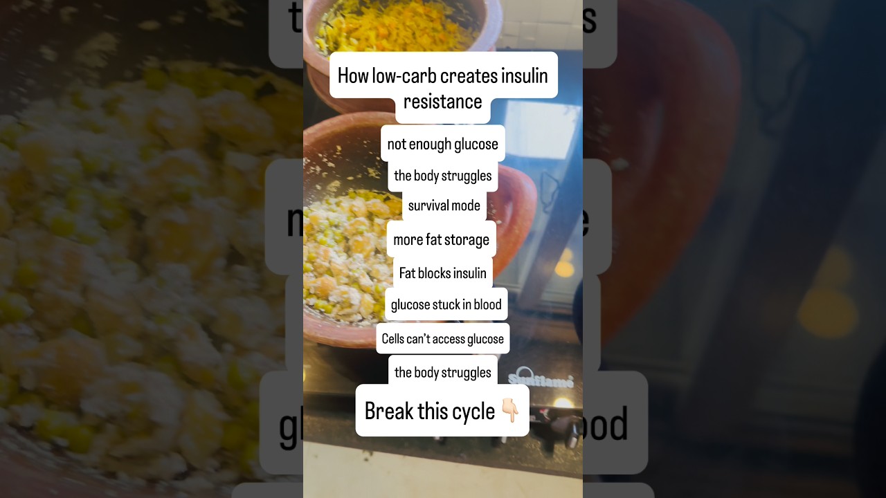
How low carb creates insulin resistance
Nidhikumari_healthcoach
1K views•2026-06-16